3-4.5.2 Fluorescence in situ hybridization (FISH)
FISH is a cytogenetic technique that uses fluorescent probes that bind to complementary targets and sample is visualized using epi-fluorescence or confocal microscopy. Using differently labeled probes, we can visualize several targets in a single sample. It is used for spatial-temporal patterns of gene expression and resolving genetic elements in chromosomal preparations.
Cells or tissues to be analyzed are fixed and permeabilized with Proteinase K to allow target accessibility. Probe is constructed and tagged using non-radioactive labels like biotin, digoxigenin or fluorescent dye (FISH). Probe must be large enough to hybridize specifically with its target. Probe is applied to fixed sample and incubated to several hours to allow hybridization. Washing is done to remove non-specific or unbound probe removal. Results are then visualized using either bright-field or confocal microscopy.
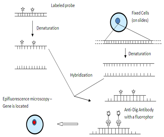
Fig3-4.5.2: Fluorescent in-situ hybridization