Mechanism of action of seven helix receptors : The seven helix receptors use trimeric GTP-binding proteins to relay signals to effector proteins inside the cells. Human genome encodes several thousand GPCR. These include receptors in the visual, olfactory (smell) and gustatory (taste) systems, neurotransmitter receptors and most of the receptors for hormones that control the metabolism of carbohydrate, amino acid and fat. GPCRs are coupled to signal-transducing trimeric G proteins. In mammals, olfactory cells alone use 500 – 1000 different seven-helix receptors to discriminate odorant molecules.
Phosphorylation of the C-terminal tail inactivates many types of seven helix receptors. Two different strategies, sometimes acting on the same receptor, provide negative feedback. One strategy is for the second messengers, which are produced in response to receptor activation to stimulate general protein kinases (including cAMP), protein kinase A (PKA) and protein kinase C (PKC) which phosphorylate the activated receptor. Phosphorylation inhibits the receptor thus allowing for crosstalk between receptors. The second strategy involves a class of protein kinases specific for the receptor themselves. They are called G-protein coupled receptor kinases. These kinases phosphorylate multiple serines or threonines on the C-terminal cytoplasmic tail of active receptors. Phosphorylation promotes binding of a regulatory protein called arrestin which inactivates the receptor by blocking interaction of the receptor with trimeric G-proteins. Arrestin binding to some seven helix receptors promotes their removal from the plasma membrane by endocytosis. G protein–coupled receptors transduce signals from extracellular hormones to associated effector proteins. In the resting state, when no ligand is bound to the receptor, the Gα subunit is bound to GDP and complexed with Gβγ . As shown in the Figure 6, ligand binding shifts the equilibrium from the resting conformation towards the active conformation. Active receptor promotes dissociation of GDP from α subunit of multiple trimeric G-proteins, allowing GTP to bind. This dissociates Gα from Gβγ, each of which activate downstream effectors that produce the second messengers cAMP and diacylglycerol (DAG) as shown in Figure 7. cAMP and DAG activates PKA and PKC, which phosphorylate active receptors on their C-terminus. This attracts arrestin, putting the receptor into the inactive adapted state.
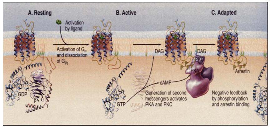
Figure 7 : Activation and adaptation of a seven helix receptor: A, resting; B, active; C, adapted.
Epinephrine Binds to Several Different G Protein–Coupled Receptors. All epinephrine receptors are G protein– coupled receptors, the different types are coupled to different G proteins. These receptors are of interest because they trigger different intracellular signal-transduction pathways. Both subtypes of β-adrenergic receptors, termed β1 and β2, are coupled to a stimulatory G protein (Gs) that activates the membrane-bound enzyme adenylyl cyclase . Once activated, adenylyl cyclase catalyzes synthesis of the second messenger cAMP. That binding of epinephrine to β-adrenergic receptors induces a rise in cAMP.
Interesting facts:
- Receptor tyrosine kinase is the first receptor to be discovered.
- G proteins were discovered by Alfred G. Gilman and Martin Rodbell when investigating stimulation of cells by adrenaline.
- Skin contains approximately 640,000 sense receptors scattered unevenly over the body's surface.