2. Nuclear receptors: Nuclear receptors are intracellular proteins expressed in the nucleus of a cell. These receptors are members of a family of proteins known as the nuclear receptor super family. Nuclear receptors constitute a superfamily of dimeric C4 zinc-finger transcription factors that bind lipid-soluble hormones and interact with specific response elements in DNA. Steroid receptors are homodimers of zinc-finger proteins that reside within the nucleus (except for the glucocorticoid receptor which resides in the cytosol until it binds its ligand). Until their ligand finds them, some steroid receptors within the nucleus associate with histone deacetylases (HDACs), keeping gene expression repressed in those regions of the chromosome.
Structure of nuclear receptors: Nuclear receptors constitute a superfamily of dimeric C4 zinc-finger transcription factors. They are modular in structure and contain the following structural domains :
N-terminal regulatory domain (A-B) : The A-B domain is highly variable in sequence between various nuclear receptors. It contains the activation function 1 ( AF-1 ) whose action is independent of the presence of ligand. The transcriptional activation of AF-1 is normally very weak but it synergizes with AF-2 in the E-domain to produce an upregulation of gene expression.
DNA-binding domain ; DBD (C) : It is a highly conserved domain containing two zinc fingers that binds to specific sequences of the DNA called hormone response elements (HRE) as shown in Figure 3.
Hinge region (D) : It is the flexible domain that connects the DBD with the LBD. It influences subcellular distribution and intracellular trafficking.
Ligand binding domain LBD (E) : Its sequence is moderately conserved but it is highly conserved in structure between the various nuclear receptors. The structure of the LBD is referred to as an alpha helical sandwich fold in which three anti parallel alpha helices (the sandwich filling) are flanked by two alpha helices on one side and three on the other (the bread). The ligand binding cavity is within the interior of the LBD and just below is present three anti parallel alpha helical sandwich filling. Along with the DBD, the LBD contributes to the dimerization interface of the receptor. In addition, it binds coactivator and corepressor proteins. The LBD also contains the activation function 2 ( AF-2 ) whose action is dependent on the presence of bound ligand.
C-terminal domain (F) : It is highly variable in sequence between various nuclear receptors.
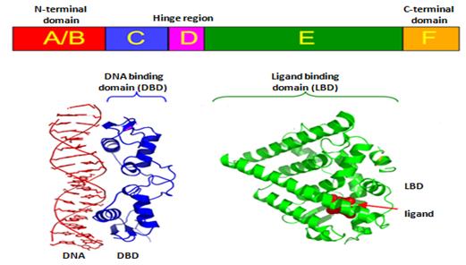
Figure 3 : Structural Organization of Nuclear Receptors (estrogen receptor)
Top – Schematic amino acid sequence of a nuclear receptor.
Bottom – 3D structures of the DBD (bound to DNA) and LBD (bound to hormone) regions of the nuclear receptor