Light Microscope: The Fluorescence microscope
The absorption and subsequent re-radiation of light by organic and inorganic specimens are typically the result of well-established physical phenomena described as being either fluorescence or phosphorescence.
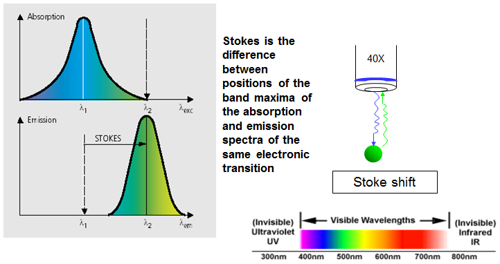
Figure 10.13: Typical absorption and emission spectra and principle of stoke shift.
• The fluorescence microscopy is an essential tool particularly in biology, the biomedical sciences and in materials science due to attributes that are not readily available in other contrast modes with traditional optical microscopy.
• In this case, the specimen is exposed to ultraviolet, violet and / or blue lights.
• The application of an array of fluorochromes has made it possible to identify cells and sub-microscopic cellular components with a high degree of specificity amid non-fluorescing material.
• This reveals bright images of the object resulting from the fluorescent light emitted by the specimen. In fact, this microscope is capable of revealing the presence of a single molecule.
• Although this cannot provide a spatial resolution below the diffraction limit of specific specimen features, the detection of fluorescing molecules below such limits is always achieved.