Light Microscope: Differential Interference Contrast Microscope
Differential interference contrast (DIC) microscopy also known as Nomarski microscopy (NM) is an optical microscopy illumination technique (see Figure 10.11) used to enhance the contrast in unstained and transparent samples. It works on the principle of interferometry to gain information about the optical path length of the sample. A relatively complex lighting scheme produces an image with the object appearing black to white on a grey background.
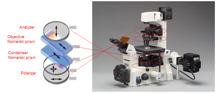
Figure 10.11: Schematic of DIC microscopy principle and set up.
This image is similar to that obtained by phase contrast microscopy but without the bright diffraction halo.
It creates an image by detecting differences in refractive indices and thickness of distinctive parts of specimen. This is an excellent way to observe living cells.
Figure 10.12 displays typical images obtained through DIC microscope.
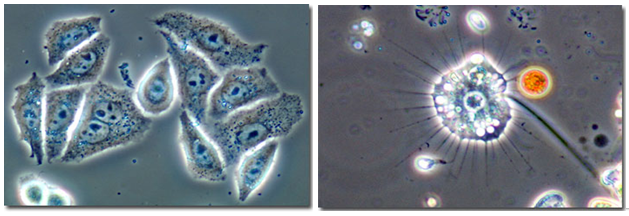
Figure 10.12: Images obtained using DIC microscope for HeLa Cell structure (left) and Heliozoans (right) [2].
Ref.[2]. http://www.microscopyu.com/staticgallery/dicphasecontrast/helapc.html.