Light Microscope: The Fluorescence microscope
Figure 10.15 displays the typical images of cells obtained using the fluorescence microscope.
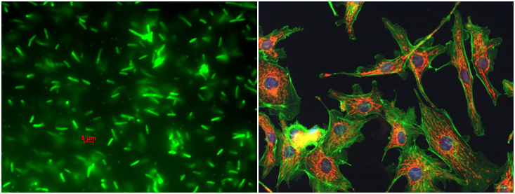
Figure 10.15: Fluorescence microscope image of RV308 cells electroporated with fluorescein labeled oligonucleotides (left) [3] and cells (right) [4].
Ref.[3]. http :// 2009.igem.org/Team:Freiburg_bioware/Project/invivo.
Ref.[4]. http://web.chem.ucsb.edu/~kalju/.
Quiz 10:
(Q10.1). When focusing a specimen, which component you start with for the adjustment?
(Q10.2). When using the high power objective, what the knobs should be used?
(Q10.3). What type of microscopes is commonly used in the most science classes?
(Q10.4). Which part of the microscope can adjust the amount of light that hits the slide?
(Q10.5). The objectives are attached to what part of the microscope (it can be rotated to click the lenses into place)
(Q10.6). A microscope has an ocular objective of 10x and a high power objective of 50x. What is this microscope's total magnification?