Optical microscopes:
Light Microscope: Phase contrast
Phase contrast microscopy is an optical microscopy technique that converts phase shifts in light passing through a transparent specimen to brightness changes in the image. Phase shifts are invisible, but become visible when shown as brightness variations.
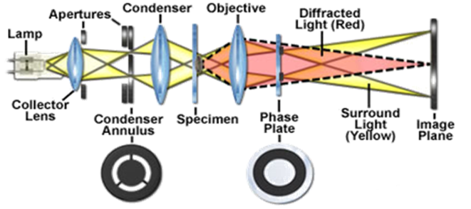
Figure 10.09: Schematic of Phase cntrast light microscope.
The basic principle to make phase changes visible in phase contrast microscopy is to separate the illuminating background light from the specimen scattered light (see Figure 10.09). This makes up the foreground details, and to manipulate these differently.
Phase contrast microscopy is particularly important in biology. It reveals many cellular structures (right image) that are not visible with a simpler bright-field microscope (left image), as shown in the Figure 10.10.
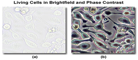
Figure 10.10: Images of Cellular structure obtained using (a) bright-field and (b) phase contrast microscopes.
• Light rays in phase produce brighter images, while light rays out of phase produce darker images.
• Contrast is created because light waves are out of phase.
• Enhances the contrast between intracellular structures having slight differences in the refractive index.
• Used to examine living organisms or specimens that would be damaged/altered by attaching them to slides or staining. Excellent way to observe living cells.
• Two types
• Phase-contrast microscope
• Differential interference contrast microscope