Optical microscopes:
Light Microscope: Bright-field
Bright-field microscopy is the simplest microscope of all the types of optical microscopes. Sample is illuminated using white light from bottom side as shown in Figure 10.04 and the image is observed from above. The contrast in the specimen is caused by absorbance of some of the transmitted light in dense areas of the sample.
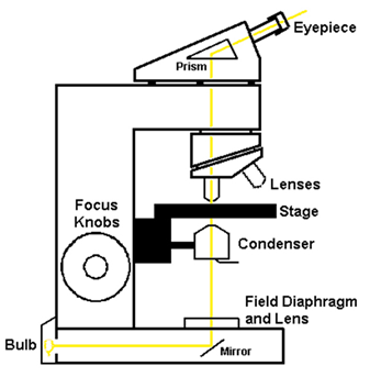
Figure 10.04: Schematic of light microscope.
The typical appearance of a bright-field microscopy image is a dark sample on a bright background and hence the name.
Series of lenses are placed for suitable magnification. It has one or few ocular lenses to view the sample image.
• Total magnification = Magnification of objective lens x Magnification of an ocular lens
• Most have the condenser lens (direct light through specimen)
• Oil immersion lens improves the light through objective lens and increases resolution, as shown in Figure 10.05. Oil immersion is mainly used to increase the resolution of a microscope. This is obtained by immersing both the objective lens and the specimen in a transparent oil of high refractive index (~ 1.515) and specific optical and viscosity characteristics. This helps to increase the numerical aperture of the objective lens.
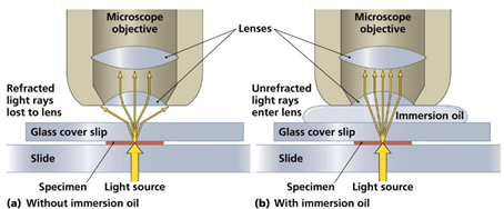
Figure 10.05: Light microscope with and without oil immersion lens.
Bright field is the most universal technique used in light microscope. Typical image observed for Hemlock leaf using the light microscope is shown in Figure 10.06.
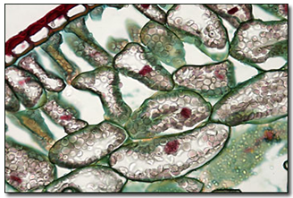
Figure 10.06: Optical image of Hemlock leaf [1].
Ref.[1]. http://micro.magnet.fsu.edu/primer/anatomy/brightfieldgallery/hemlockleaf40xlarge.html.