Procedure:
Preparing diphenylalanine nanotubes
- Weigh 5 mg of diphenylalanine peptide and dissolve it in 50 μl HFIP. This gives a peptide concentration of 100 mg/ml.
- Dilute the peptide into distilled water to a final concentration of 2 mg/ml.
- Allow the solution to age for one day at room temperature. This results in the self-assembly of the peptide to give tubular assemblies that can be visually observed.
AFM sample preparation
- Take a small (~1 cm2 area) V1 quality mica sheet.
- Place the mica piece on a solid support and peel off the upper mica surface using a
sticky
tape to obtain a smooth surface.
- Deposit ~0.1 -1 μg of the peptide on the mica surface.
a. Take 2 μl of the clear part of the diphenylalanine solution in a microfuge tube and add 198 μl distilled water.
b. Take 50 μl (theoretical peptide amount ~1 μg) of the peptide solution and deposit on the freshly cleaved mica surface.
- Remove the excess fluid from the mica surface after one minute. This can be done by carefully touching lint-free tissue paper to the edge of mica.
- Air-dry the mica.
AFM imaging
We shall be discussing the steps that are to be followed for performing the intermittent mode (also known as AC mode) imaging on the AFM from Agilent Technologies (Figure 36.4).
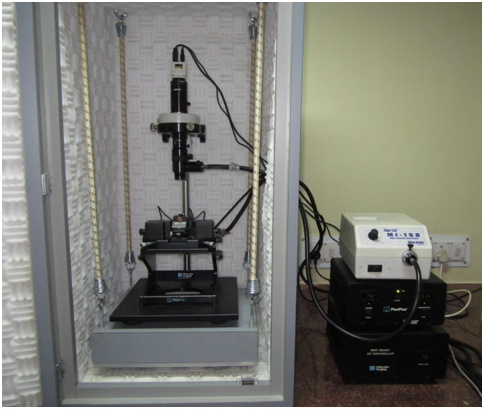
Figure 36.4 An atomic force microscope (Agilent Technologies)