Modes of AFM
Figure 36.2 shows the Lennard-Jones potential for a pair of atoms.
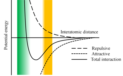
Figure 36.2: Lennard-Jones potential energy curve for two atoms
An AFM experiment can be performed in either attractive or repulsive regime of the Lennard-Jones potential. Depending on the working regime of the Lennard Jones potential, AFM imaging methods are divided into three basic modes:
Contact mode AFM: In contact mode AFM, the probe is brought in contact with the specimen surface; the interaction between the probe and the specimen, therefore is repulsive. As the tip is in contact with the sample, the frictional forces are very high during scanning. Contact mode imaging, therefore, may not be suitable for soft samples including biological samples.
Non-contact mode AFM: In non-contact mode AFM, a cantilever with very high spring constant is oscillated close to its resonance frequency. The probe does not contact the specimen and interacts with it through long range surface interactions. The forces between the probe and the specimen are very small, of the order of piconewtons. This mode, therefore, is well-suited for soft samples; resolution, however, is compromised.Intermittent mode or tapping mode AFM: A stiff cantilever is oscillated close to its resonance frequency, at a probe-specimen separation that allows a small part of oscillation lie in the repulsive regime of the Lennard-Jones potential. The probe-specimen interaction therefore varies from long-range attraction to weak repulsion. The tip intermittently touches the sample while scanning. Interaction of the probe with the sample surface causes changes in the amplitude and the phase of oscillation. This mode of imaging allows imaging with very high resolution and has become the method of choice for scanning the soft biological samples.
In this experiment, we shall be using intermittent mode of imaging to study the ordered superstructures formed by a self-assembling peptide. In intermittent contact mode of imaging, user can define an amplitude set-point. This amplitude can be maintained using a feedback mechanism that moves the cantilever up or down to maintain the user defined vibrational amplitude. The cantilever displacement directly corresponds to the height of the specimen. In constant amplitude mode, the oscillating cantilever scans the sample, moving up/down to maintain the defined amplitude. A plot of cantilever displacement against the specimen coordinates generates the topographic image of the specimen surface.