Atomic Force Microscopy
Aim:
To study the nanotubes formed by diphenylalanine using atomic force microscopy
Introduction:
Atomic force microscopy belongs to the class of microscopic methods together known as scanning probe microscopy (SPM). The working principle of scanning probe microscopes is very different from conventional optical microscopes. An SPM scans the surface of the sample using a very fine pointed probe measuring one or more of the sample properties at each point. Atomic force microscope (AFM) is a scanning probe microscope that measures the force between the the probe and the specimen.
An AFM has a cantilever (a cantilever is a beam fixed at only one end) that has a finely pointed probe, also referred to as the AFM tip, at its free end. The other end is anchored to a piezoelectric displacement actuator (Figure 36.1).
Attachment to the piezoelectric material allows precise positioning of the cantilever with respect to the specimen. For imaging, the probe is brought in close proximity to the specimen surface. Interaction (attractive or repulsive) between the probe and the specimen imposes a bending moment on the cantilever. Responding to this moment, the cantilever deflects towards or away from the specimen. The deflection of cantilever is detected using a laser beam that is focused on the cantilever, just above the probe. The back surface of the cantilever is highly reflective, the reflected beam is focused on a split photodiode (Figure 36.1). The cantilever is scanned across the specimen surface in a raster pattern. Any deflection in the cantilever as a result of sample interaction causes displacement in the laser spot on the photodiode; this displacement signal (difference in response in the upper and lower sectors of the split diode) is used to calculate the deflection in the cantilever.
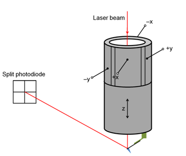
Figure 36.1: Diagrammatic representation of a cantilever attached to a piezoelectric tube. The laser beam falls on the back of the cantilever and gets reflected to hit the split photodiode detector.