In repeated subcultures on the proliferation medium, the embryogenic cells continue to multiply without the appearance of embryos. However, if the PEMs are transferred to a medium with a very low level of auxin (0.01-0.1 mgl-1 ) or no auxin in the medium (embryo development medium ; ED medium), they develop into embryos. The presence of an auxin in the proliferation medium seems essential for the tissue to develop embryos in the ED medium. The tissues maintained continuously in auxin-free medium would not form embryos. Therefore, the proliferation medium is called the ‘induction medium' for SE and each PEMs as an unorganized embryo.
Cytokinin : There are reports of somatic embryo induction and development in cytokinin containing medium, but these reports are very few compared to those reporting induction by auxin alone or auxin plus cytokinin. Cytokinin, in general, induced SE directly without the callusing of explant. In most cases, TDZ is used as cytokinin, a herbicide, which mimics both auxin and cytokinin effects on growth and differentiation. The other cytokinins are also used when zygotic embryos are used as the explant source. The most commonly used cytokinins are BAP and Zeatin.
In Azadirachta indica , somatic embryo differentiation was influenced by the culture medium as well as the stage of embryo at culture. Maximum somatic embryogenesis occurred directly from the explant on BAP containing medium when early dicotyledonous stage of embryos were cultured. Medium with 2,4-D induced only neomorph differentiation directly from the explant. While torpedo shaped embryos showed both neomorph formation as well as somatic embryogenesis on BAP containing medium (Figure 8.6).
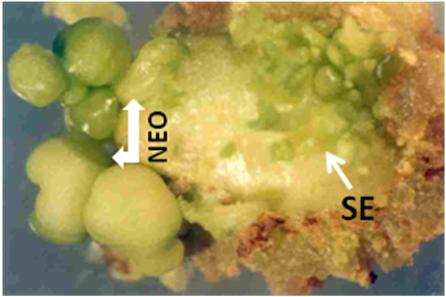
Figure 8.6: An explant showing differentiation of neomorphs (NEO) and somatic embryos (SE) on the same explant
Neomorphs were suppressed embryos with green, smooth, shiny surface and solid interior (Figure 8.7A). Although they were epidermal in origin like somatic embryos with heart shape notch but showed monopolar germination and no clear cut radicular region (Figure 8.7B).
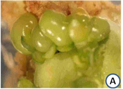
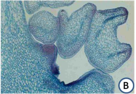
Figure 8.7: A. An explant showing direct differentiation of neomorphs. Some of these structures also show cotyledon-like flaps. The portion of the explant in contact with the medium has proliferated into a brownish green callus
B. A histological section of A, showing epidermal origin of a neomorph of various shapes. It has a well differentiated epidermis and compactly arranged internal cells. These structures are loosely attached to the explant and show provascular strands.