SEM: Interaction with sample
• Upon impinging on the specimen, the primary electrons decelerate and in losing energy transfer it inelastically to other atomic electrons and to the lattice. Through continuous random scattering events as shown in Figure 17.05, the primary beam effectively spreads and fills a teardrop-shaped interaction volume with a multitude of electronic excitations.
• Secondary electrons: Common imaging mode relies on detection of these electrons originating from a subsurface depth of no larger than several angstroms. The signal is captured by a detector consisting of a scintillator -photomultiplier combination, and the output serves to modulate the intensity of a CRT. The image magnification is the ratio of scan lengths on the CRT to that on the specimen.
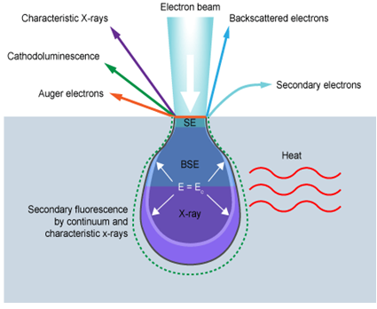
Figure 17.05: Schematic of electron interacting with specimen surface [5].
• Backscattered electrons: These are the high energy electrons that are elastically scattered and essentially possess the same energy as the incident electrons. The probability increases with the atomic number Z of the samples. Although the identification of element is not feasible from such information, the contrast can develop between regions of the specimen that differ widely in Z.
Ref.[5]. http://www.ammrf.org.au/myscope/sem/background/concepts/interactions.php#detail.