SEM: Principles
• SEM has an electron column as shown in Figure 17.02, where the electrons are generated and arranged through electric and magnetic fields in a proper direction to achieve the required incident beam at the sample surface.
• The whole process is operated under vacuum to avoid collision between electrons and gas molecules.
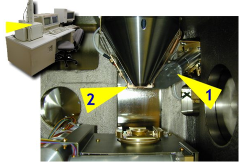
Figure 17.02: Photoview of interior of SEM [2].
When the electron beam hits the sample, the interaction of the beam electrons from the filament and the sample atoms generates a variety of signals.
Hence, the SEM has several detectors to view the electron signals from the sample. Two of these detectors can be seen from inside the specimen chamber (see figure 17.02). The secondary electron detector used for detecting the secondary electrons looks like a Faraday cage (1). The backscattered electron detector is located above the sample (2) to detect backscattered electrons.
• Unlike the light microscope (in which light forms an instant real image of the object), the electrons in an SEM do not form a real image.
• SEM scans its electron beam line by line over the sample by using a set of scan coils. This is similar to using a flashlight in a dark room to scan the room from one side to another side.
• Subsequently, the image is built on a monitor (cathode ray tube (CRT)).
• The interaction between the electron and specimen is discussed in next slide.
Ref.[2]. http://academic.udayton.edu/ShirleyWright/SEM/Principle