Atomic force microscopy
An atomic force microscope (AFM), like SEM, provides information about the surface properties of the specimen. The resolution of the images is determined by the tip shape and diameter as described in the previous lecture (Figure 19.6). With AFM, resolution comparable to or even better than TEM is routinely achieved. A big plus of AFM over EM is its potential to perform imaging of the liquid samples as well, albeit with lesser resolutions (~20 – 50 nm). Imaging of liquid samples is one of the most desired characteristics of biological microscopy. Soft biological samples are easily analyzed using intermittent mode/tapping mode AFM. Furthermore, an AFM analysis does not require tedious sample preparation. Let us go through some of the applications AFM has been utilized for:
Imaging of dry samples: The specimen is deposited on an atomically-smooth substrate, typically mica and dried. The dried specimen is directly studied by AFM without requiring any staining. The ability to provide resolutions comparable to TEM makes AFM a powerful tool in nanotechnology. Figure 20.5 shows a tapping mode AFM image of a self-assembled peptide.
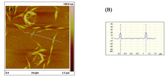
Figure 20.5 A height-mode AFM image of a self-assembled peptide (A). Height of the fibers indicated by blue crosses in panel A (B).
Cell biology: Owing to its ability to operate on liquid samples, AFM has been used to study the real-time biological processes. Migrating epithelial cells, dynamics of membrane invaginations, conformational changes in membrane proteins, and assembly/disassembly of structural proteins have been studied in real time using AFM.
Nucleic acid research: AFM has slowly emerged as a powerful tool to analyze the structures of the nucleic acids and the various processes they are involved in. Three-way and four-way DNA junctions have been analyzed using AFM. Time-lapse AFM imaging has been used to study the mechanism of branch migration in the four-way DNA junctions. Molecular processes like DNA replication, transcription, translation, and DNA- protein interactions have been studied using time-resolved AFM imaging. Exploiting their highly-specific assembly, nucleic acids have been designed to obtain ordered self-assembled structures that have been characterized using AFM.