(vi)Histopathology: Histopathology is the area of pathology that deals with the anatomical changes in the tissues. The tissue samples are sliced into thin sections and stained with a dye. A number of stains are available and the choice of stain depends on the histological features one needs to study. For example, hematoxylin and eosin stain is a routinely used stain to study the morphological features of tissue samples, congo red is often used to identify the amyloid plaques, Giemsa stain is used for identifying the parasites such as plasmodium. If a fluorescent stain is used, the specimens can be analyzed by fluorescence microscopy.
(vii) Cytopathology: Cytopathology, as the name suggests, is the study of pathological conditions at the cellular level. Any change in the cellular morphology or anatomy following an infection, as a result of a metabolic disorder, or a cellular condition like sickle cell anemia can be studied by staining the cells and analyzing them using any of the light microscopic methods.
(viii) Cellular membranes and intracellular structures: A cellular feature can be selectively labeled using fluorescently labeled antibodies (immunofluorescence, discussed in lecture 15) or the fluorescent dyes that selectively bind to the cellular structures. For example, probes that specifically bind to the cellular organelles like nucleus, mitochondria, and lysosomes are commercially available. e.g. DAPI for DNA staining.
(ix) Membrane proteins: Fluorescently labeled membrane proteins can be studied using total internal reflection fluorescence (TIRF) microscopy as discussed in lecture 15. TIRF allows selective excitation of the fluorophores that are in close proximity with the sample substrate such as a glass slide.
(x) Live cell imaging: Inverted microscopes allow direct microscopy of the cultured cells.
(xi) Protein dynamics and localization: Green fluorescent protein (GFP) and its variants have made it possible to selectively label the proteins within a cell. Live cell imaging using fluorescence microscopy allows studying the dynamics and localization of the proteins in the cells.
(xii) Co-localization of the proteins: Confocal laser scanning microscopy (CLSM) scans a specimen and gives the plot of intensity in the two dimensional coordinate space. Performing scanning experiments in closely spaced focal planes provides the three dimensional distribution of the fluorophore inside the cell. This allows to study if two proteins are close together within the cell. Figure 20.1 shows the confocal images recorded for two proteins; protein A is labeled with the green fluorescent protein (GFP) while protein B is labeled with the RFP. The co-localization of the red emitting protein with the green emitting protein gives yellow color (Figure 20.1D)
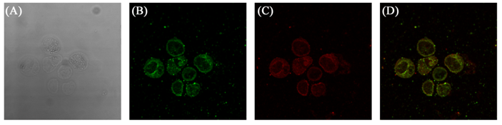
Figure 20.1 A bright-field image of a cell expressing protein A-GFP and protein B-RFP (A); a confocal image recorded for GFP (B); a confocal image recorded for RFP (C); a superimposed image showing co-localization of the two proteins (D).