Nanotomography: A TEM micrograph is the two-dimensional projection of a three dimensional object. Recording a large number of images at different tilt angles, however, can be used to construct the three-dimensional model of the specimen as shown in figure 20.3.

Figure 20.3 Tomography: a diagrammatic representation of a cylinder's images recorded at different angles.
Cryoelectron microscopy: We have seen that the specimens to be analyzed by TEM as well as SEM need to be completely dehydrated. Imaging under hydrated conditions is a highly desirable feature for the imaging of biological specimens. Cryoelectron microscopy (Cryo-EM) is a TEM method that makes it possible to analyze the specimen under hydrated conditions. Cryo-EM has become a major tool for determining the structures of large biomolecular complexes that are difficult to study by routine structure determination methods such as X-ray crystallography and NMR spectroscopy. The biomolecule is dissolved in a suitable buffer that stabilizes its native structure. A small amount of the sample is placed on the EM grid and excess sample is removed using blotting paper. The sample coated grid is plunged into a cryogen; the rapid cooling inhibits formation of ice crystals that could damage the specimen. The specimen therefore is in the amorphous ice. The frozen specimen is studied under TEM. As no staining is done, the contrast of cryo-EM is very poor. As the specimen, prior to freezing, is isotropic, the images are obtained for all possible orientations of the molecules. The resolution can be enhanced by stacking the images of the molecules captured in the same orientation e.g. a and b in figure 20.4. The images for these molecules can be cut out, aligned, and stacked one over another. Noise being random gets cancelled out giving a better contrast.
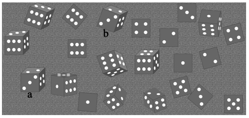
Figure 20.4 Images of the identical dice in different orientations. Image ‘a' can be aligned with image ‘b' by rotating it 20° (clockwise) and translating it to the coordinates of image ‘b'. Stacking of a large number of such images is used to enhance contrast in cryo-EM.