Nucleotides: Nicotinamide adenine dinucleotide in its reduced form, NADH and the flavin adenine dinucleotide in its oxidized form, FAD are fluorescent in the visible region of the electromagnetic spectrum. It is not necessary for all the biomolecules to have an intrinsic fluorophore to perform fluorescence experiments. Fluorescent groups can be covalently incorporated into the molecules making them fluorescent with desirable fluorophore. Such externally incorporated fluorophores are called extrinsic fluorophores.
Applications of fluorescence
Protein folding: High sensitivity of tryptophan fluorescence to the polarity of solvent makes it an interesting intrinsic fluorescent probe for studying protein folding. In the proteins having Phe and Tyr, Trp can be selectively excited at 295 nm. In water and other aqueous solutions, tryptophan fluoresces with an emission maximum, λmax around 350 nm. A tryptophan present in the hydrophobic environment usually displays a blue shift in the emission spectrum and an increase in quantum yield. . Due to the hydrophobic nature of the indole side chain, tryptophans are usually buried inside the core of the proteins. The folding can therefore be studied by monitoring the Trp fluorescence as protein folds burying the water-exposed Trp residues inside the protein.
Peptide-lipid interactions: Interaction of the peptides having Trp residues with lipid bilayers can easily be studied using fluorescence spectroscopy. Interaction of the peptide with lipids brings the tryptophan in relatively hydrophobic environment causing a blue shift in emission spectrum (Figure 7.1).
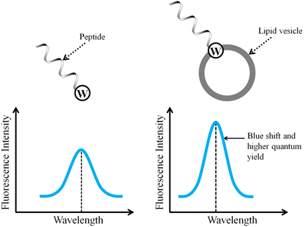
Figure 7.1 Spectral changes in tryptophan fluorescence upon binding to lipid bilayers