Biological fluorophores
Amino acids: Aromatic amino acids tryptophan (Trp), tyrosine (Tyr), and phenylalanine (Phe) are perhaps the most important intrinsic biological fluorophores. Proteins harboring these amino acids become intrinsically fluorescent.
Proteins: Proteins are fluorescent due to the presence of aromatic amino acids that fluoresce in the near UV region. Certain proteins, however, do fluoresce in the visible region. Green fluorescent protein (GFP), for example, fluoresces in the green region of the electromagnetic spectrum. The discovery of green fluorescent protein has revolutionized the area of cell biology research. It is therefore important to see what green fluorescent protein is and why it fluoresces in the visible region (See Box 1).
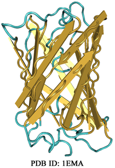
Box 7.1: Green Fluorescent Protein (GFP) Green fluorescent protein, abbreviated as GFP was discovered by Shimomura and coworkers in 1962. The protein was isolated from the jellyfish, Aequorea victoria, that glows in the dark. GFP is a 238 amino acid long protein that folds into an 11-stranded β-barrel structure wherein an α-helix passes through the barrel.
The fluorophore of the GFP, p-hydroxybenylideneimidazolinone is formed by the residues 65-67 (Ser-Tyr-Gly) and is present in the α-helix passing through the barrel. The excitation spectrum of GFP exhibits a strong absorption band at 395 nm and a weak band at 475 nm. Emission is observed at ~504 nm i.e. in the green region. GFP is an excellent fluorophore with a molar absorption coefficient of ~30000 M-1 cm-1 at 395 nm and fluorescence quantum yield of 0.79. GFP has been engineered through extensive mutations to remove the undesirable properties that could affect its use as a potential fluorophore. For example, a Ser65 → Thr65 mutant has improved quantum yield and its major excitation band shifted to 490 nm. GFP has the tendency to form oligomers, seriously questioning its use as a fluorescent probe. The aggregation tendency has also been removed through extensive mutations. GFP can easily be tagged to a protein by expressing the fusing gene (GFP gene fused with the gene expressing the desired protein). The GFP then acts as a reporter for all the processes the linked protein is involved in. Several color variants of GFP have been generated through modifications in the residues that constitute the fluorophore. Development of the GFP variants with varying excitation and emission characteristics has made it possible to label the proteins differentially. This is a huge breakthrough and allows easy monitoring of the biological processes using fluorescence microscopy as discussed in lectures 15 and 20. |