Applications
- Identification of functional groups: As has already been discussed, IR spectroscopy allows identification of functional groups. Carbonyl (C=O) is an interesting functional group worth discussing. Carbonyl is a double bond (high spring constant, k) with very high polarity. Stretching vibration of carbonyl group causes large changes in the dipole moment consequently resulting in a very intense absorption band. Furthermore, the frequency of carbonyl stretching does not differ significantly for aldehydes, ketone, carboxylic acids, and esters (Table 10.1). The large intensity and relatively unchanged frequency of carbonyl stretching allows easy identification of the carbonyl compounds (It is important to note that carbonyl stretching frequency can be much lower for amides and much higher for anhydrides and acid chlorides).
- Identification of compounds: The fingerprint region of the IR spectrum is unique to each compound. It is possible to identify a compound from its IR spectrum if the spectrum for the compound is already known and available for comparison. This is particularly useful in pharmaceutical research and development. A patented drug, if suspected to be synthesized by another pharmaceutical company, can easily be identified by comparing the IR spectra in the fingerprint region.
- Presence of impurities: Comparison of the IR spectra of the given compound with the spectra of pure compound helps in the assessment of its purity. It is important to ascertain the purity of the active molecule and the excipients used in preparing drug formulations.
- Structural transitions in lipids: Structural lipids are those that are organized in bilayers in biological membranes. Glycerophospholipids constitute the major class of the structural lipids (Figure 10.7). The lipids have several structural phases such as a gel phase with all-trans conformation and a liquid crystalline phase where gauche conformations are also present. Methylene (–CH2–) stretching vibrations give the most intense absorption band in lipids as expected for a molecule having long hydrocarbon chains.
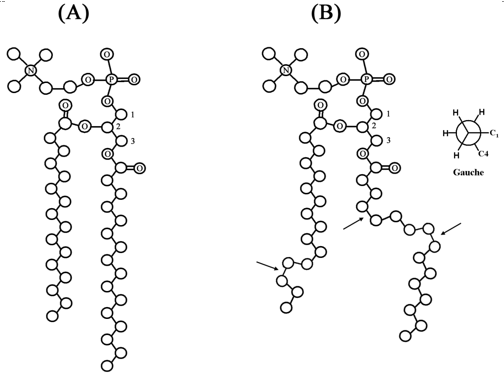
Figure 10.7. Structure of a glycerophospholipid: all-trans conformation (A); lipid with trans and gauche conformations (B), gauche conformations are indicated with arrows
Both –CH2– stretching and bending vibrations are sensitive to the conformations of the lipids and therefore provide information about the transition of lipids between different phases. Vibration modes of the head group and the interfacial region also provide useful information about local acyl chain conformation. Carbonyl stretching vibration (1750 – 1700 cm-1) in the ester bond is sensitive to the conformation of the local acyl chain conformation.
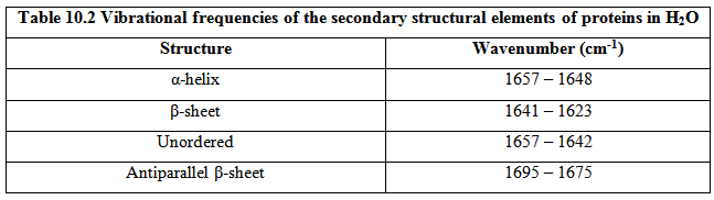
- Protein and peptide structure: Infrared spectroscopy is routinely used to study the structures of proteins and peptides. Like CD spectroscopy, the region of interest in determining the conformation of the polypeptide backbone is the peptide bond. The peptide group results in nine distinct bands, labeled as amide A, B, and I-VII. Amide I is the most useful band in studying the polypeptide backbone conformation. Amide I band (1700 – 1600 cm-1) arises largely due to the carbonyl stretching with small components of C–N stretching and N–H bending. The frequency of carbonyl stretching vibration is sensitive to the H-bonding, and therefore to the conformation of polypeptide backbone. The frequencies of absorption of different secondary structural elements are shown in Table 10.2