37.4 |
Microscopic methods of characterization
|
|
Even microscopic methods make use of more or less the same principles, but these methods are generated by bombarding the surface with high-energy electrons, which are then multiplied several times so as to produce a magnified image of the surface on the fluorescent screen.
Few methods like STM, AFM work based on quantum mechanical principles. |
|
This method takes advantage of the wave nature of light to attain higher magnification. It has nothing to do with the electron microscopy principles, only the magnification is comparable. In SEM a beam of electrons is generated in vacuum which is collimated by electromagnetic condenser lenses and scanned across the sample surface by an electromagnetic deflection coil. Primary imaging is done by collecting secondary electrons that are released by the sample. Then, the secondary electrons are magnified and finally made to fall on fluorescent screen to obtain final image. |
|
Instrumentation and working: |
1. |
Electron source: The electron source in SEM is an electron gun, a tungsten wire bent in ‘V’ shape, and applied |
|
voltage is as high as 10 keV, which then produces electrons from the tip. This whole set up is also called Wehnelt cylinder. The electrons so produced are accelerated and converged by applying anodic potential at a slight distance from the Wehnelt apparatus. |
|
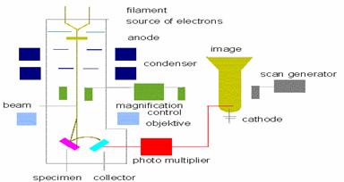
Figure 37.4 Block diagram of Scanning Electron Microscope |
2. |
Electromagnetic lenses: Unlike in typical microscopes, SEM electromagnetic lenses are used to collimate the |
|
electron beam. Here, a magnetic field is used to perform the job of condenser and objective lenses which finally focuses the beam on the sample. |
3. |
Deflection coils: The role of deflection coil is simply to scan the sample surface, which is mounted to sample |
|
platform by shifting the beam position. This is accomplished by applying positive potential to these coils. Normally solid sample can be glued directly on the platform, but powdered samples should be made into paste and then tapped on platform. |
4. |
Secondary Electron detector: Secondary electrons are generated from the surface after colliding the surface |
|
with high-energy electron beam. These are received by a net like secondary electron detector, to which a positive potential of magnitude 10 keV is applied to grab the slightest emission of secondary electrons. Since strength of secondary electrons from the surface is very low, it is necessary to apply such large positive potential. |