Atomic Force Microscope: Working principle
Eye of nanotechnology
• It allows researchers to observe and manipulate molecular and atomic level features.
• It works by bringing a cantilever tip in contact with the surface to be imaged as shown in Figure 19.02. An ionic repulsive force from the surface applied to the tip bends the cantilever upwards. The amount of bending, measured by a laser spot reflected on to a split photo detector, can be used to calculate the force. By keeping the force constant while scanning the tip across the surface, the vertical movement of the tip follows the surface profile and is recorded as the surface topography by the AFM.
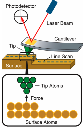
Figure 19.02: Schematic of AFM working principle.
• The predecessor of AFM is Scanning Tunneling Microscopy (STM) or the Scanning Tunneling Microscope. It was invented in 1981 by G. Binnig and H. Rohrer who shared the 1986 Nobel Prize in Physics for their invention. An excellent technique, STM is limited to imaging conducting surfaces.
• AFM has much broader potential and application because it can be used for imaging any conducting or non-conducting surface.
• In many fields (life science, materials science, electrochemistry, polymer science, biophysics, nanotechnology, and biotechnology) of nanoscience and nanotechnology, it provides the ability to view and understand events as they occur at the molecular level which will increase our understanding of how systems work and lead to new discoveries in many fields.
• AFM has a number of advantages over other techniques making it a favorite among leading researchers.
• It provides high-resolution and three-dimensional information in real space with little sample preparation involving low-cost. In-situ observations, imaging in fluids, temperature and environmental controls are all available.