Bright-field imaging mode:
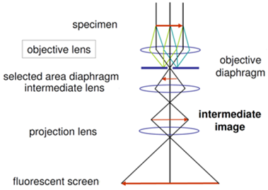
Figure 14.01: Schematic ray diagram of bright-field imaging mode.
The most common mode of operation for a TEM is the bright field imaging mode. In this mode the contrast formation is formed directly by occlusion and absorption of electrons in the sample. Thicker regions of the sample, or regions with a higher atomic number will appear dark, whilst regions with no sample in the beam path will appear bright – hence the term "bright-field".
This mode is obtained by intentionally excluding all diffracted beams and only allowing the central beam passing through the specimen as shown in the Figure 14.01.