Transmission Electron Microscope:
Instrumental Part:
Objective Lens:
• The objective lens (see Figure 12.06) forms an inverted initial image, which is subsequently magnified. In addition, in the back focal plane of the objective lens a diffraction pattern is formed. Hence, the objective aperture has to be utilized to select the components. Note that the objective lens usually would not provide a magnification of more than 50✗ and any magnification of the image is mainly done by using projector lenses.
Projector Lens:
• Magnification in the electron microscope can be varied from few hundreds to several hundred thousands of times.
• This is achieved by varying the strength of the projector lenses and intermediate lenses as shown in Figure 12.06.
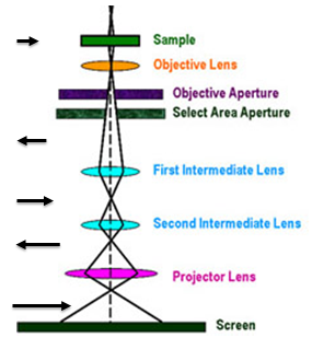
Figure 12.06: Schematic of lens system including projector lens in TEM.
• Note that all lenses need not (unnecessarily) be used at lower magnifications.
• The introduction of each lens in the system for magnification changes the direction of final image (see arrow mark on the left side) on a fluorescent screen.