A transmission electron microscope has the following components along its optical path:
- Electron source
- Condenser lens
- Specimen stage
- Objective lens and projector lens
- Screen/photographic film/CCD camera
In the electron gun, electrons are emitted from the surface of the cathode and accelerated towards the anode by high voltage (Vo) to form a high energy electron beam. All lenses in the electron microscope are electromagnetic. Charged electrons interact with the magnetic fields and magnetic force deflects or focuses an electron beam. The condenser lens system controls the beam diameter and convergence angles of the beam incident on a specimen.
Sample preparation
Preparation of specimens is the most tedious step in TEM. To make the material electronically transparent material thickness is limited. Specimens have to be prepared with thickness of ~ 100 nm. For higher atomic weight material, the specimen has to be thinner. TEM sample preparation depends on type of materials used.
For bulk material TEM specimen preparation is done in two steps :
- Pre-thinning : thinning to 0.1 mm thickness
- Final-thinning: thinning to 100 nm thickness
Pre-thinning: In pre-thinning stage specimen less than 1mm thick is prepared by mechanical cutting (with a diamond saw). Then 3-mm-diameter disc is cut using punch before further reduction of thickness. Grinding is most commonly used to reduce the thickness of metal and ceramic specimens
Final thinning: Final thinning is mainly done by electrolytic thinning & ion milling, which create a dimpled area on pre-thinned specimens that have regions of electron transparency as shown in Fig.2. In the electrolytic thinning, metal specimen is made the anode in an electrolytic cell. On passing current the metal is gradually dissolved and deposited on the cathode. Finally tiny holes appear, the edge of which are suitable for electron transparency. In ion milling method samples are bombarded with beam of energetic ions to reduce the thickness by knocking atoms out of the specimen. This method can also be used for ceramics and other non-conducting materials. For polymeric and biological specimens ultramicrotomy method is used where specimen is cut into thin sections by cutting tool such as glass knife or diamond knife.
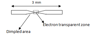
Fig. 2 . Thinning of bulk specimen for TEM analysis
For powder samples, such as catalysts, the powder size is reduced by grinding, to a very fine size till the particle thickness is small enough to allow electron transmission. These fine particles are then suspended in volatile solvent such as isopropanol. A drop of this particle suspension is placed on thin carbon foil supported by a conventional microscope grid. On evaporation of solvents the powder particles are ready for the observation. Alternatively the powders can be embedded in some suitable matrix such as epoxy resin or metals, from which a flat sheet of 3 mm diameter is cut out to study by conventional methods.
Magnification and resolution
Magnification and resolution is defined in the same way as transmission light microscope.
- Magnification of any convergent lens M

f = focal length of lens ; v = distance between image and lens
- Resolution of electron microscope can be written as

α = Angle through which beams are deflected ; λ = wave length of electrons
For λ =0.0037 nm (wave length of 100 kV electrons ) and α = 0.1 radians (~5 degrees) resolution of ~0.02 nm is obtained