Electron Microscopy
Microscope forms an enlarged image of the original object in order to convey its internal or external structure. The magnification and resolution of microscope is given as
(a) Magnification = objective lens × eyepiece lens
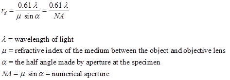
(b) Resolution (rd) is defined as closest spacing of two points which can be clearly seen through the microscope as separate entities. Resolution can be determined as:
By increasing the dimension or by employing large number of lenses, the magnification can be increased, while shorter wavelength yields higher resolution.
Electrons are considered as radiation with wavelength in the range 0.001 - 0.01 nm compared to 400- 700 nm wavelength of visible light used in an optical microscope. In an electron microscope, a focused electron beam is used instead of light to examine objects, and the image of the specimen is obtained on a very fine scale. Because of the use of large number of lenses and electrons of very low wavelength, magnification and resolution for electron microscopes is much higher. High magnification and resolution makes electron microscopes extremely useful for revealing ultrafine details of material microstructure. Optical microscopes have a maximum magnification power of 1000, and resolution of 0.2 μm compared to resolving power of the electron microscope that can reach 1,000,000 times and resolution of 0.2 nm. Hence, electron microscopes deliver a more detailed and clear image compared to optical microscopes.
- Two main types of electron microscopes
- Transmission electron microscopes (TEM)
- Scanning electron microscopes (SEM)
Transmission Electron Microscopes (TEM)
The optics of the TEM is similar to conventional transmission light microscope. It was developed in 1930s. It is capable of displaying magnified image of thin specimen with magnification in range of 103 to 106 . Information that can be obtained using TEM include :
- Topography: surface features, texture
- Morphology: shape and size of the particles
- Crystallographic arrangement of atoms
- Composition: elements and the their relative amounts
Interaction of electron beam with sample
Interaction of electron beam with the sample result is three types of electrons:
- Unscattered electrons: These are electrons that are transmitted through the thin specimen without any interaction with the sample. Transmission of unscattered electrons is inversely proportional to the specimen thickness. The areas of specimen that are thicker will have fewer transmitted unscattered electrons and will appear darker. Conversely thinner areas will have more transmitted electrons and will appear lighter.
- Elastic scattered electrons: These are incident electrons that are scattered by the atoms of the specimen in elastic fashion that is without any loss of energy of electrons. The elastically scattered electrons form diffraction pattern that yield information about the orientation, atomic arrangements and phases present in the area being examined.
- Inelastic scattered electrons: Incident electrons that interact with the specimen atoms in inelastic fashion, lose energy during the interaction fall in this catagory. The extent of loss of energy by incident electrons depends on the characteristic of interacting elements and is used to study the compositional and bonding (i.e. oxidation state) informations of specimen region being examined.
Working Principle and Instrumentation
The Fig. 1 shows the arrangement of components for transmission electron microscope. An electron gun at the top of the microscope emits electrons that travel through vacuum in the microscope column. Vacuum is essential to prevent strong scattering of electrons by gases. Electromagnetic condenser lenses focus the electrons into a very thin beam. Electron beam then travels through the specimen and then through the electromagnetic objective lenses. At the bottom of the microscope, unscattered electrons hit the fluorescent screen giving image of specimen with its different parts displayed in varied darkness, according to their density. The image can be studied directly, photographed or digitally recorded.
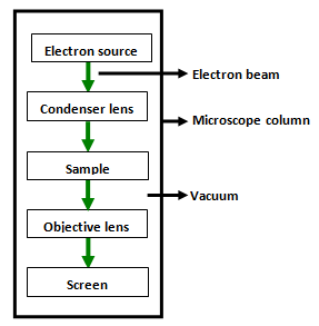
Fig. 1. Schematic diagram illustrating transmission electron microscope