Lab Experiment 30.1: Calculate the IC50 of chloroquinine against malaria parasite in an in-vitro microscopic schinzonticidal assay.
Background Information: Light microscope can be visualized the object in two different modes (bright field/dark field) and both of these modes give different information of an object. Both of these modes are extensively been used to perform multiple task in the biological research.
Bright-field microscopy: In a bright-field microscope, both diffracted (diffracted by the specimen) and undiffracted (light that transmits through the sample undeviated) lights are collected by the objective lens (Figure 30.3). The image of the specimen is therefore generated against a bright background, hence the name bright-field microscopy. Most biological samples are intrinsically transparent to the light resulting in poor contrast. To increase the contrast of the image, the specimens are therefore generally stained with the dyes.
Dark-field microscopy : Dark-field microscopy increases the contrast of the image by eliminating the undiffracted light. If there is no specimen in the optics path, no light is collected by the objective lens. Presence of specimen results in the diffraction of light; the objective lens collects the diffracted light generating a bright image against a dark background.
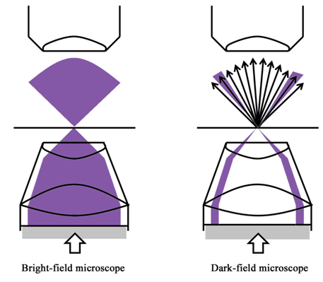
Figure 30.3: Optical diagrams of bright-field and dark-field microscopes
Material and Instruments:
1. RPMI 1640 cell culture media
2. Albumax-II
3. 0.22µm membrane filter
4. Filtration Unit
5. Autoclave
6. Vacuum Pump
7. Upright microscope