Light Microscopy
Introduction: Light microscopy is the simplest form of microscopy. It has tools that are used to observe the small organisms or object and even macromolecules. It has wide variety of microscopic tools for studying the biomolecules and biological processes.. It includes all forms of microscopic methods that use electromagnetic radiation to achieve magnification.
Instrumentation of a typical light microscope-The typical diagram of a light microscope is given in the Figure 31.1. The light is produced by a lamp (with tungeston filament) as source and light rays are focused on the specimen by the condenser. The specimen is kept on the stage and firmed by clipped present on the side. The light diffracted by the sample is then collected by the objective lens (objective lens varies from 10x-100x magnification) and additional magnification is achieved by the eyepiece (usually gives additional 10x magnification). Hence, if you observe a sample with 40x objective lens, microscope is actually magnifying the object by 400x (40x from objective and 10x from the eye piece, 40x10=400x).
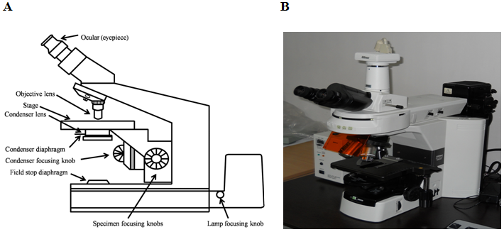
Figure 31.1: Instrumentation of a typical light (binocular) microscope with its different components. (A) Schematic Diagram and (B) Actual microscope
Light microscopes come in two designs: upright and inverted (Figure 31.2).
Upright microscope: In an upright microscope, the objective turret is usually fixed and the image is focused by moving the sample stage up and down.
Inverted microscope: In an inverted microscope, the sample stage is fixed and objective turret is moved up and down to focus the final image.
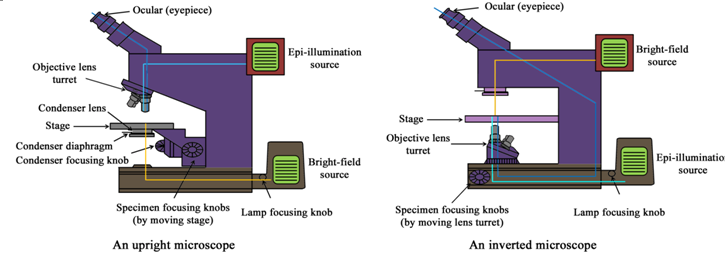
Figure 31.2 Designs of upright (A) and inverted (B) microscopes