Aim:
To record the far-UV circular dichroism spectrum of a protein
Introduction:
Circular dichroism, abbreviated as CD, is a chiroptical spectroscopic tool that is routinely employed to study the secondary structural elements of proteins and peptides. The technique is also used to determine the conformational stability of the proteins as discussed in the previous lecture, study the kinetics of folding/unfolding, binding with ligands, and to determine if an expressed, purified protein is correctly folded. Unlike X-ray crystallography and NMR spectroscopy, that can provide residue specific structural information, circular dichroism provides the overall secondary structural components with no residue-specific information. The advantages of CD include small sample requirement, rapid measurements, and measurements under physiological conditions. Protein/peptide concentration of ≤ 20 μg/ml is usually sufficient for recording spectra in far-UV region; furthermore, recording is usually complete within 5-10 minutes.
Let us now see what actually circular dichroism is? Dichroism literally means “two colours”. In chiroptical spectroscopy, dichroism refers to the differential absorption of lights with different polarizations. Circular dichroism therefore refers to the differential absorption of lights with different circular polarizations. You may already be familiar with plane or linearly polarized light. Let us see what circular polarization is and how it is achieved. Consider two plane polarized electromagnetic waves of same wavelength, polarized in two perpendicular planes and out of phase by 90°. The 90° phase difference implies that when one of the waves has maximum amplitude, the other one has zero amplitude (Figure 9.1).
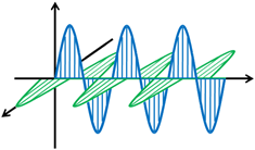
Figure 9.1 Two plane polarized waves (blue and green), polarized in two perpendicular planes and out of phase by 90º. Only electric field vectors are shown here for clarity. |
The superposition of these two plane waves is shown in figure 9.2.
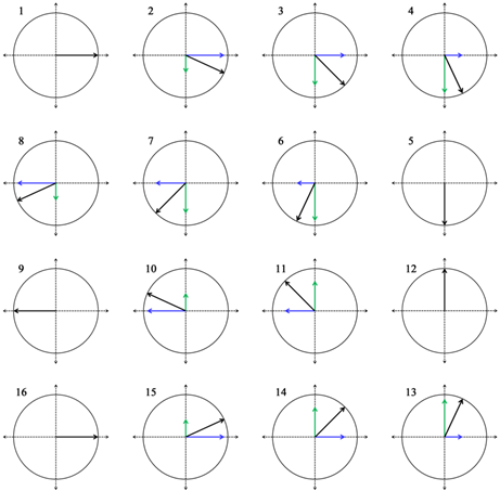
Figure 9.1 Generation of circularly polarized light through superposition of two plane polarized waves (blue and green), polarized in two perpendicular planes and out of phase by 90º. Only electric field vectors are shown here for clarity. |