Let us have a look at the CD spectra characteristic of the different structural components of the proteins (Figure 9.2).
- α-helix: The right handed α-helix displays two negative absorption bands centered around 222 nm (n → π* transition) and 208 nm (a part of the π → π* transition) and a strong positive band around 192 nm (a part of the π → π* transition).
- β-sheet: β-sheets are characterized by the presence of a negative band centered around 216-218 nm (n → π* transition) and a positive band of comparable intensity at around 195 nm (π → π* transition).
- β-turn: A β-turn comprises of a four residue protein motif that causes the polypeptide backbone to take an approximately 180° turn. The CD spectrum for a β-turn is not well defined. A typical β-turn, however, shows a weak negative band around 225 nm (n → π* transition), a strong positive band between 200 – 205 nm (π → π* transition), and a strong negative band (π → π* transition) between 180 – 190 nm.
- Random coil: Random coil or unordered conformation shows a weak positive band around 218 nm (n → π* transition) and a strong negative band (π → π* transition) below 200 nm.
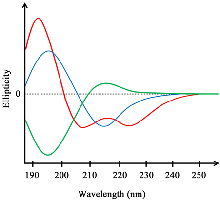
Figure 9.2 Far UV circular dichroism spectra of α-helix (red), β-sheet (blue), and unordered conformation (green) |
The CD spectrum of a protein can be written as a linear combination of the spectra of all the structural components:
CD (protein) = a CD(α-helix) + b CD(β-sheet) + c CD (Random coil)
As the CD spectra of different structural components are quite distinct, it is possible to estimate the fraction of different structural components in a protein from its CD spectrum. As discussed in lecture 5, proteins also have chromophores that absorb in the near UV region. These include the aromatic amino acids and disulfide linkages. The CD of aromatic amino acids is highly dependent on their environment and therefore near UV CD of proteins can provide the information about the environments these residues reside in as well as their orientations in the structure. As it provides information about the tertiary region, near UV CD is also referred to as tertiary CD in the context of the proteins.