| The role microscopy plays in understanding biological systems and processes hardly needs any introduction. The first uses of microscopes for observing the biological specimens date back to | |
| 1660s. It would have not been possible to identify and understand the organization of microorganisms without using microscopy. Light microscopy is used to identify the microorganisms based on their morphology and the specific stains they take up. A routine quantitative application of microscopy is to count the number of different cells per unit | 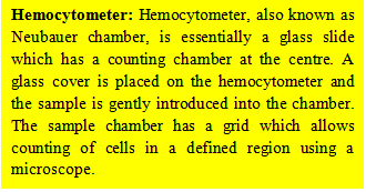 |
| volume of blood or any other sample using a hemocytometer. Presence of cells that are not expected in the healthy individuals may be an indicator of anomaly/disease. For example, a simple microscopic analysis of blood sample will identify the sickle cell anemia; presence of pus cells in urine, quantified by microscopy, is an indicator of infection. Light microscopy uses light as the illumination radiation and is perhaps the most familiar form of microscopy. In the simplest microscopic methods, a specimen is illuminated by visible light and observed either against a bright background (bright-field microscopy) or a dark background (dark-field microscopy). Fluorescence microscopy, one of the most commonly used microscopic methods in biological research, has emerged as a very powerful tool for studying molecular processes owing largely to the advancement in optics and discovery of the green fluorescent protein and development of its analogs with different spectral properties (discussed in lectures 15 and 16). Confocal laser scanning microscopy (CLSM) is a type of fluorescence microscopy that allows imaging of the samples at different focal planes i.e. light emitting from below or above the desired focal plane is eliminated. This results in very high lateral resolution and allows determining the spatial localization of the molecules (discussed in lecture 16). Total internal reflection fluorescence (TIRF) microscopy is another type of fluorescence microscopy wherein the optics allows imaging of the molecules that are in close proximity to the microscopic slide (discussed in lecture 15). The resolution of light microscopes depends on the wavelength of the light used. The smaller the wavelength of the light used, the better the resolution obtained. Wavelength of the visible light imposes a resolution limit of ~0.2 μm on the light microscopes (discussed in lecture 14). What it means is that the two point objects closer than ~0.2 μm cannot be resolved used a light microscope |
|