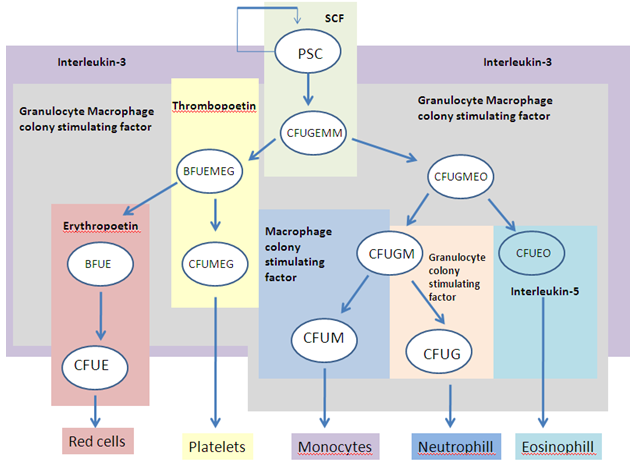
Figure 2: Schematic representation of the role of growth factors in normal haemopoiesis. PSC, pluripotential stem cell; SCF, stem cell factor. For other abbreviations see Figure 1.
Erythropoiesis
Erythropoiesis is the name for the process which leads to the formation of red blood cells (RBCs) or more properly termed as the erythrocytes. The normal life span of RBC’s is about 120 days, thus new erythrocytes need to be formed. The overall process occurs in five days and the bone marrow is the site for the production of RBCs. A condition known as hypoxia which is shortage in RBC’s oxygen carrying capacity leads to the release of the growth factor erythropoietin. Other growth factors which are released are IL-1, IL-4, IL-6, IL-11, IL-12, and SCF. Furthermore, Insulin, Growth hormone, and steroid hormones are very crucial in RBC production. EPO acts on precursor RBC cells which are Burst Forming Unit-Erythroid (BFUE) and Colony Forming Unit-Erythroid cells (CFUE) leading to their proliferation. The scheme given below summarizes the process of erythropoiesis. During sudden hypoxia due to massive blood loss the entire aforesaid process takes place in three days.
↓ O2 tension → ↑ EPO → ↑ RBC’s precursors (BFU-E and CFU-E) → ↑ differentiation & proliferation → ↑ mature RBC’s release in 5 days.
They are six morphologically identifiable stages in erythroid differentiation which can be visualized under the microscope using Romanowsky (or Geimsa) stained slides. The different stages are namely:
a. Pronormoblasts: These cells makes up about 1-2% of all nucleated cells in the bone marrow. The cytoplasm is very basophilic, i.e., has very dark blue color.
b. Basophilic normoblasts: These cells constitutes up to 4% of all nucleated cells in the bone marrow. Under the microscope the cytoplasm shows deep blue color.
c. Polychromatophilic normoblasts: These cells makes up to 10-20% of all nucleated cells in the bone marrow. The cytoplasm varies in color due to the synthesis of hemoglobin, which leads to a wide range of colors consisting of a mixture of gray, blue, mauve, and/or violet.
d. Orthochromic normoblasts: The cytoplasm of these cells has a resultant color of pale grayish-blue-violet due to the presence of hemoglobin
e. Reticulocytes: The retics appear slightly larger than normal erythrocytes, with a varying degree of color. The cytoplasm may be irregular and might have inclusions known as “basophilic stippling”, which are the residual RNA remaining in the cells.
f. The mature erythrocyte (RBC): The erythrocyte has a diameter of about 7μ and width of about 2μ. The cell lacks nucleus, and mitochondria.