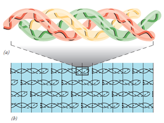
Figure 2: The structure of collagen I. (a) The monomer of collagen. (b) Collagen I molecules become aligned in and a bundle of collagen I molecules, such as that shown here, form a collagen fibril.
This figure has been adapted from Cell and Molecular Biology Concepts and Experiments by Karp, 2010.
Polysaccharides of Matrix
The structural proteins of the extracellular matrix are rooted in polysaccharides called glycosaminoglycans (GAGs). One sugar of the disaccharide is either N-acetylglucosamine or N-acetylgalactosamine and the second is usually glucuronic acid or iduronic acid. They can also be sulfated like the chondroitin sulfate, dermatan sulfate, heparan sulfate, and keratan sulfate. These polysaccharides are highly negative in charge and bind positively charged ions and water molecules to form hydrated gels. The function of such gels is to provide support to the matrix. Hyaluronan is the only GAG that occurs as a single long polysaccharide chain. GAGs also attach with proteins through Serine residues and are known as proteoglycans. A number of proteoglycans interact with hyaluronan to form large complexes in the extracellular matrix e.g., aggrecan which is the major protein of the cartilage. Proteoglycans also interact with collagen and other matrix proteins to form gel-like networks in which the fibrous structural proteins of the extracellular matrix remain rooted.
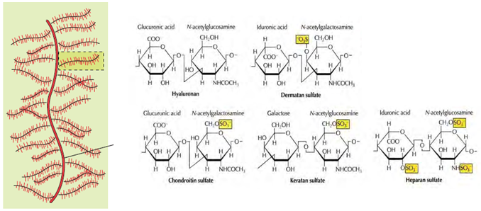 Figure 3: Proteoglycan complex with the major types of matrix glucosaminoglycans. Glycosaminoglycans consist of repeating disaccharide units. With the exception of hyaluronan, the sugars frequently contain sulfate. The figure has been adapted from “ The Cell, A Molecular Approach” by Geoffrey M. Cooper, 4 th Ed. 2007.