Electron Microscopy-II
Lab Experiment 35.1 : Localize the protein inside the macrophage using transmission electron microscope.
Background Information: In the transmission electron microscope; the electrons were focused on a thin specimen and the electrons transmitted through the specimen were detected. Figure 35.1 shows a simplified optical diagram comparing a light microscope with a transmission electron microscope.
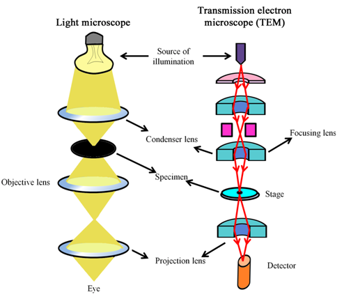
Figure 35.1: A simplified comparison of optics in a light microscope with that in a TEM.
Transmission electron microscopes usually have thermionic emission guns and electrons are accelerated anywhere between 40 – 200 kV potential. However, TEM with >1000 kV acceleration potentials have been developed for obtaining higher resolutions. Owing to their brightness and very fine electron beams, field emission guns are becoming more popular as the electron guns.
Material and Equipments
1. Paraformaldehyde
2. Glutaraldehyde
3. PBS (1X)
4. 1% Tween-20
5. BSA (Fat free, acetylated): Prepare 2% BSA solution in PBS and filter with the 0.45mm filter to rmove particulate matter.
6. Primary antibody (anti-protein): An antibody can be developed against protein (antigen of interest) in rabbit or mice.
7. Secondary antibody: An antibody coupled with gold particle and directed against mouse IgG.
8. Uranyl acetate
9. Osmium tertaoxide
10. Propylene oxide
11. Ethanol
12. Epon resin
13. LR white resin
14. Microtome
15. Transmission Electron microscope.