Structure of Prokaryotic cells- A prokaryotic cell is much simpler and smaller than eukarotic cells. It lacks membrane bound organelles including nucleus. A typical prokaryotic cells is shown in Figure 3.1, A. The description of different structural feature of prokaryotic cells is as follows-
1. Outer Flagella: A flagellum attached to the bacterial capsule is a central feature of most of the prokaryotic cell especially of the motile bacteria. It provides motion or locomotion to the bacteria and be responsible for chemotaxsis of bacteria. Movement of bacteria towards a chemical gradient (such as glucose) is known as chemotaxsis. Flagellum is a part of cell wall and its motion is regulated by motor proteins present inside the cell. Flagellar motion is an energy consuming process and it is governed by an ATPase present at the bottom of the shaft. It is made up of protein flagellin and reduction or suppression of flagellar protein reduces bacterial infectivity (pathogenicity) and ability to grow.
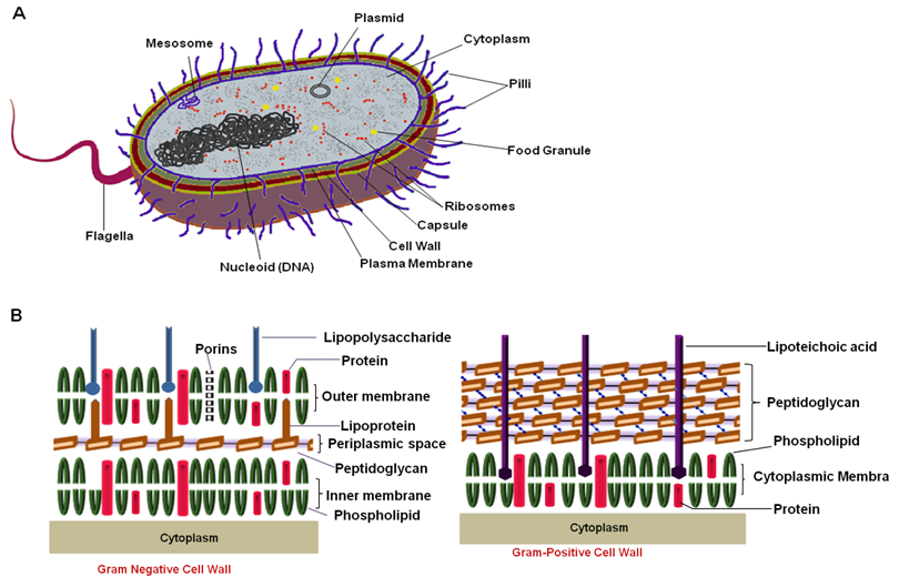
Figure 3.1: Structural details of a typical prokaryotic cell. (A) Whole cell and (B) composition of cell wall of gram negative and positive bacteria.
2. Bacterial surface layers: Bacteria posses 3 anatomical barriers to protect the cells from external damage. Bacterial capsule is the outer most layer and made up of high molecular weight polysaccharides. It is impermeable to the water or other aqueous solvent and it is responsible for antigenicity of bacterial cells. Cell wall in bacteria and its response to gram staining is the basis of classification of bacterial species.
| WHAT IS GRAM STAINING? Gram staining is developed by a Danish scientist Hans Christian Gram. This technique differentiates bacterial strains based on their cell wall composition, especially thickness of peptidoglycan layer. A detail staining procedure is given in following paper ( Use of the gram stain in microbiology. Beveridge, TJ (2001) Biotech Histochem 76 (3): 111–8. Pubmed ID: 11475313 ). During the staining procedure bacterial sample is stained with two dyes, crystal violet and safarin . During a washing step with non-polar solvents such as alcohol or acetone (decolorization), gram –ve bacteria leave the blue stain due to a thin peptidoglycan layer in cell wall whereas gram +ve bacteria retains both stains and appear as Pink . |