Force mode AFM/Force spectroscopy: Force mode of AFM is not an imaging mode. A typical force spectroscopy experiment is schematically shown in Figure 19.5. Briefly, the sample is brought close to the cantilever, pushed against it causing deflections in it, and then withdrawn. A plot of force (depends on the spring constant of the cantilever) against the distance is called a force spectrum. Force spectroscopy mode is often used to study the interactions of the tip with the sample and to determine the mechanical properties of the specimen.
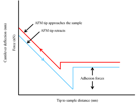
Figure 19.5 A diagrammatic representation of typical approach and retract force spectra.
Resolution
Atomic force microscopes can provide resolutions comparable to that obtained with electron microscopes. As neither light nor particles are used to generate the images, resolution of atomic force microscopes does not depend on any wavelength. The resolution of an AFM is determined by the shape and the diameter of the tip. Figure 19.6 shows what influence the tip diameter has on the resolution in an AFM. It is also evident that the resolution in the X-Y plane is poorer as compared to that in the Z-direction . A Z-resolution of ~0.2 nm or better is often achieved using AFM.
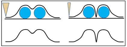
Figure 19.6 Effect of tip diameter on the lateral resolution of an AFM.
Advantages of AFM
Both AFM and EM provide very high resolution images but AFM has few distinct advantages over EM:
- Easy sample preparation: AFM does not involve a tedious sample preparation. A sample to be analyzed can simply be placed on a smooth surface and scanned.
- Imaging in solution: Unlike EM; it is possible, in fact routine; to record AFM images in solution. No other microscopic method, except the scanning probe microscopes, provides a sub-nanometer resolution in solution.
- Manipulation: An AFM tip can be used to mechanically manipulate the specimen at very high spatial resolution.