Two photon and multiphoton laser scanning microscopy
If a fluorophore absorbs the light of energy, ![]() where λ is the
wavelength
of the absorbed radiation; it is possible to excite the fluorophore with the light of wavelength 2λ if two photons are simultaneously absorbed by the molecule (Figure 16.5).
where λ is the
wavelength
of the absorbed radiation; it is possible to excite the fluorophore with the light of wavelength 2λ if two photons are simultaneously absorbed by the molecule (Figure 16.5).
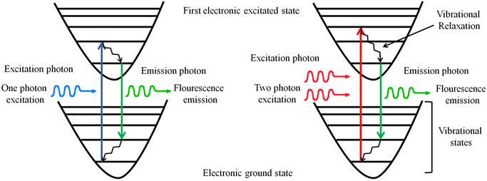 Figure 16.5 A simplified Jablonski diagram showing single-photon and two-photon excitation of a fluorophore.
The probability of simultaneous absorption of two photons is very small; multiphoton microscopes therefore need very intense light sources. Pulsed infrared lasers, however, have realized the multiphoton microscopy. Titanium:sapphire lasers operating at 800 nm can cause excitation of the fluorophores with λmax ~ 400 nm through two photon absorption. Multiphoton fluorescence microscopy offers following advantages over single photon microscopy:
- Biological specimens absorb the near-IR radiation very poorly as compared to the UV and blue green radiation, the electromagenetic region commonly used for fluorescence microscopy; this implies that a thicker specimen can be studied using multiphoton microscopy.
- As the fluorophores are excited at ~2λ in a two photon fluorescence imaging experiment, the incident and the emitted radiations are well separated; this separation allows detection of the emitted radiation clear of the excitation radiation and the Raman scattering.
- The probability of simultaneous absorption of two photons depends on the square of the light intensity. The laser light in a two-photon set up does not excite the fluorophores along its path due to insufficient photon density to cause two-photon absorption. A photon density high enough to cause excitation is achieved only at the focus, thereby exciting the molecules only in the focal plane (Figure 16.6). A multiphoton microscope therefore does not require a pinhole for recording confocal images.
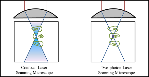
Figure 16.6 A comparison of the excitation region in a confocal laser scanning microscope and a two photon laser scanning microscope.