We have seen how total internal reflection fluorescence (TIRF) microscopy eliminates the light from most of the sample except the thin layer of the sample in contact with the sample slide. An intrinsic limitation of the TIRF microscopy is that the thin layer that can be studied is always fixed. It would be interesting if any thin layer within the specimen could be studied; this would allow localization of the molecules within the cell. Laser scanning confocal microscopy does exactly that. Figure 16.2 shows how a small modification in a fluorescence microscope allows collection of fluorescence from a thin section of the sample. Including a pinhole before the eyepiece rejects the light coming from most of the sample; the light is collected only from a thin section of the sample resulting in a sharp image (Figure 16.2B). This rejection of out-of-focus light by using a pinhole is the principle behind confocal microscopy
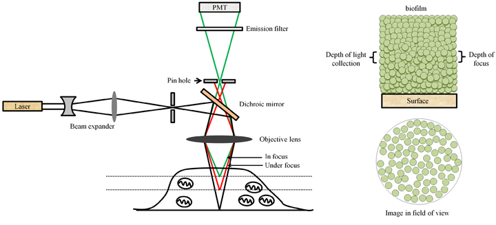 Figure 16.2 Optical diagram of a confocal laser scanning microscope; the pinhole rejects the light coming from non-confocal planes (A); a hypothetical image generated from the light coming from the focal plane. Compare the image with that shown in figure 16.1.