It is possible to put the GFP (or its variant) tag at either ends of the protein. This is important for labeling the proteins that have localization signals at the N-terminus; N-terminal labeling of such proteins would abolish their proper localization.
Fluorescence microscope
Figure 15.2 shows the optical diagram of an epifluorescence microscope, perhaps the simplest of all fluorescence microscopes. In an epifluorescence microscope, the illumination of the specimen as well as the collection of the fluorescence light is achieved by a single lens. This has become possible due to the incorporation of dichroic mirror in the optics. A dichroic mirror is largely reflective for the light below a threshold wavelength and transmissive for the light above that wavelength.
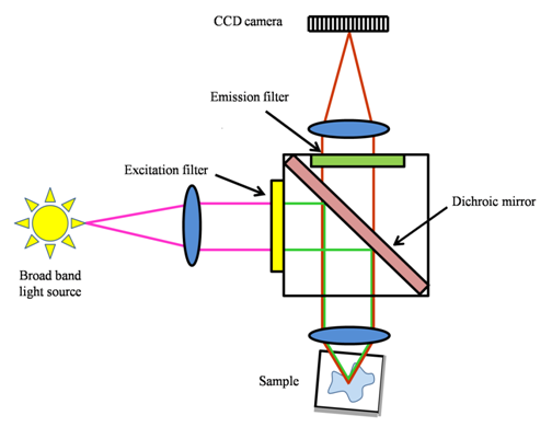
Figure 15.2 A diagram showing the optical path in an epifluorescence microscope.
The microscope has a high power lamp source, usually a mercury or xenon arc lamp. An excitation filter transmits the band of the excitation radiation. The excitation radiation is reflected by the dichroic mirror towards the condenser/objective lens that focuses the light on the specimen. Light emitted by the fluorescent molecules (higher wavelength due to Stokes shift) is collected by the same lens and is transmitted by the dichroic mirror towards the ocular lens. Figure 15.3 shows a comparison between a brightfield and a fluorescence image of the Cos-7 cells expressing GFP.
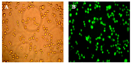
Figure 15.3 Bright-field (A) and epifluorescence (B) images of Cos-7 cells expressing GFP.