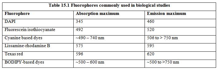
Fluorescence microscopy has come a long way since the application of fluorescence in microscopic studies in early 20th century. Unlike the other types of light microscopy that need special optics to enhance the contrast (see lecture 14), fluorescence in visible region of electromagnetic radiation is directly detected. Most biomolecules, however, are not fluorescent in the visible region. The cellular features, however, can be studied using extrinsic fluorescent probes that can go inside the cell and bind to the intracellular molecules with high specificity. Table 15.1 lists some of the fluorescent molecules routinely used for fluorescence microscopy with biological specimens. The fluorescence emission of the dyes used in biological microscopy span the entire visible region of the electromagnetic spectrum.
Immunofluorescence, that makes use of the very high specificity of antibodies towards their targets, is a very useful method for studying cellular markers and organelles. Immunofluorescence microscopic analysis of cell surface markers is straightforward wherein the cells are treated with the fluorescently labeled antibodies and studied under microscope. For intracellular targets, however, the cells are usually fixed and permeabilized to allow the antibodies enter the cells. Fluorescence microscopic analysis of cells provides information about the distribution of the target molecules in the cell. The need of fixing and permeabilizing the cells puts a restriction on immunofluorescence to be used for studying the live cells. An alternative approach is to use small fluorescent dyes that can translocate across the biological membrane and bind to the cellular targets with high specificity. Another approach includes directly labeling the molecule under study with a fluorescent tag. Carboxyfluorescein, for example, is covalently linked to the N-terminus of the synthetic peptides for performing microscopic studies. This approach, however, may not be suitable for labeling the specific molecules inside a cell. Discovery of green fluorescent protein (GFP) and developments of its variants with different spectral properties has made it possible to selectively label the proteins inside the cell using molecular cloning (strategy shown in figure 15.1)
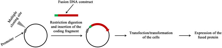
Figure 15.1 Strategy for selectively labeling a protein in a cell. The cDNA for the protein under study is fused with that of cDNA of GFP or any of its variants. The fusion DNA construct is then overexpressed in the cell.