Phase contrast microscopy
A phase contrast microscope provides very high contrast as compared to the bright-field and dark-field microscopic methods. The image in a phase contrast microscope is generated from both diffracted and
undiffracted lights
as shown in Figure 14.7. Like dark-field microscopy, the specimen is illuminated by the light coming from a ring, called a condenser annulus. The diffracted and the undiffracted lights are separated in space allowing selective manipulation of their phases and intensities. The diffracted as well as the undiffracted light is collected by the objective lens. A phase plate is placed at the back side of the objective lens that increases the phase of the undiffracted light by ![]() and decreases that of diffracted light by
and decreases that of diffracted light by ![]() as shown in Figure 14.7. A total phase difference of
as shown in Figure 14.7. A total phase difference of ![]() is therefore obtained between the diffracted and the undiffracted light beams before they are focused on the image plane. As the intensity of the undiffracted light is very high, it is selectively reduced to ~30% of the initial intensity by
a semi-transparent
metallic film on the phase plate. Two waves that have
is therefore obtained between the diffracted and the undiffracted light beams before they are focused on the image plane. As the intensity of the undiffracted light is very high, it is selectively reduced to ~30% of the initial intensity by
a semi-transparent
metallic film on the phase plate. Two waves that have ![]() phase difference interfere destructively thereby diminishing the light intensity. Any phase change caused by the specimen is therefore converted into an amplitude signal by a phase contrast microscope thereby increasing the contrast.
phase difference interfere destructively thereby diminishing the light intensity. Any phase change caused by the specimen is therefore converted into an amplitude signal by a phase contrast microscope thereby increasing the contrast.
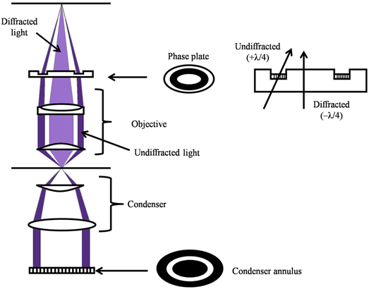
Figure 14.7 Optical diagram of a phase contrast microscope