Bright-field microscopy
In a bright-field microscope, both diffracted (diffracted by the specimen) and undiffracted (light that transmits through the sample undeviated) lights are collected by the objective lens (Figure 14.6). The image of the specimen is therefore generated against a bright background, hence the name bright-field microscopy. Most biological samples are intrinsically transparent to the light resulting in poor contrast. To increase the contrast of the image, the specimens are therefore generally stained with the dyes. However, intrinsically colored samples such as erythrocytes can directly be observed using bright-field microscopy.
Dark-field microscopy
Dark-field microscopy increases the contrast of the image by eliminating the undiffracted light. The specimen is illuminated by the light coming from a ring at an oblique angle (Figure 14.6). If there is no specimen in the optics path, no light is collected by the objective lens. Presence of specimen results in the diffraction of light; the objective lens collects the diffracted light generating a bright image against a dark background.
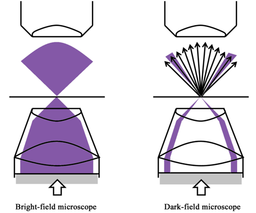
Figure 14.6 Optical diagrams of bright-field and dark-field microscopes