As is clear from the definition of resolution, lower dmin implies higher resolution. Resolution of a light microscope operating at the blue end of the visible spectrum will therefore be higher than that operating at the red end, assuming all other parameters remain same. The theoretical limit for dmin for a light microscope operating in high refractive index (typically, nmax = 1.4 for the oil used in microscopy) is ~ 0.17 μm (Assuming λ = 400 nm and sin α = 1). It is therefore an intrinsic limitation of a light microscope to resolve the particles closer than ~0.17 μm. It is evident that the resolution can be increased if the wavelength of the source radiation is reduced.
Parts of a light microscope
Figure 14.5 shows the diagram of a light microscope. The light is produced by a lamp source and focused on the specimen by the condenser. The light diffracted by the sample is then collected by the objective lens that generates a real magnified image as shown in Figure 14.3 . This image is further magnified by the eyepiece.
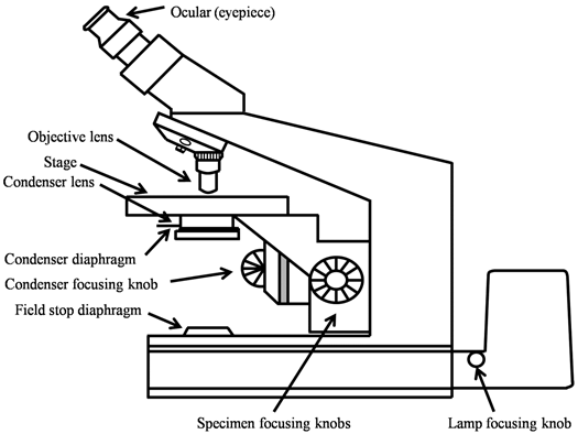
Figure 14.5 Schematic diagram of a compound microscope showing its different components