3. Transmission Electron Microscope (TEM)
Transmission electron microscopy (TEM) is a microscopic technique in which a beam of electrons is transmitted through an ultra thin specimen, resulting in interaction with the specimen as it passes through. An image is produced from the interaction of the electrons transmitted through the specimen, which is magnified and focused onto an imaging device, such as a fluorescent screen, on a layer of photographic film, or detected by a sensor. TEM has been exploited most widely in the examination of the internal surface of cells (Figure 18.3).
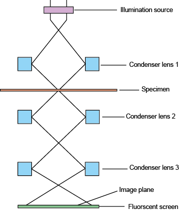
Figure 18.3: Organization of Transmission Electron Microscope
3.1. Operation of TEM
To operate TEM, it requires an ultra high vacuum and a high voltage. Through a sequence of adjustments of focus and brightness of the beam, the setting of the microscope is adjusted so that by shifting, the sample holder finds the thin area of the sample. Then tilting of the sample begins by rotating the holder. This is a way to view as much areas as we can, so we can obtain as much information.
Various images are obtained in TEM by properly utilizing the apertures and by using different types of electrons. Diffraction patterns are seen because of the scattered electrons. If the unscattered beam is selected, the bright field image is obtained. Dark field images are achieved if diffracted beams are selected by the objective aperture.
In transmission microscopy, the specimen's structure and its atomic columns are observed, thus compositional and crystallographic information is attained. However, being a very expensive technique, expertise is needed and the sample preparation phase is too difficult in order to achieve very thin samples.