1.3. Specimen preparation for electron microscopy
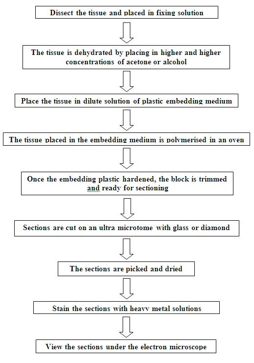
Tissues to be examined in the electron microscope must be fixed, embedded and sectioned. Fixation of tissues for electron microscopy (Scheme 18.1) is much more critical than for light microscopy because the sections are subjected to a much greater scrutiny. A fixative must stop the life of a cell without significantly altering the structure of that cell and cell components. To obtain the most rapid fixation and the least cellular damage, very small pieces of tissues are fixed and embedded. Fixatives are chemicals that denature and precipitate cellular macromolecules. Once the tissue is fixed, the water is removed by dehydration in alcohol, and the tissue spaces are filled with a material that supports tissue sectioning.
Tissues to be sectioned for electron microscopy are usually embedded in epoxy resins, such as Epon or Araldite. Sections are cut by slowly bringing the plastic block down across an extremely sharp cutting edge made of cut glass or a finely polished diamond after which the sections are thoroughly dried and stained using heavy metal solutions, such as uranyl acetate or lead citrate. These heavy metals bind to the macromolecules and provide the atomic density required to scatter the electron beams.
Scheme 18.1: SCHEMATIC REPRESENTATION OF SPECIMEN PREPARATION FOR
ELECTRON MICROSCOPY