2. Scanning Electron Microscope (SEM)
A scanning electron microscope (SEM) is a type of electron microscope that forms images of a sample by scanning it with a high-energy beam of electrons. The electrons interact with the atoms that make up the sample resulting in signal production that contains information about the sample's surface topography, composition, and other properties, such as electrical conductivity. SEM is used primarily to examine the surfaces of objects (Figure 18.2).
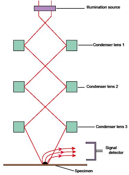
Figure 18.2: Configuration of scanning electron microscope
2.1. Operation of SEM
In SEM, a source of electrons is focussed in vacuum into a fine probe that is raster over the surface of the specimen. The electron beam passes through scan coils and objective lens that deflect horizontally and vertically so that the beam scans the surface of the sample. As the electrons penetrate the surface, numerous interactions occur that results in the emission of electrons or photons from or through the surface. A reasonable fraction of the electrons emitted can be gathered by suitable detectors, and the output can be used to modulate the brightness of a cathode ray tube (CRT) whose x- and y- inputs are driven in synchronism with the x-y voltages raster the electron beam. In this way an image is produced on the CRT. Every point that the beam strikes on the sample is mapped directly onto a corresponding point on the screen. As a result, the magnification system is simple and linear magnification is calculated by the equation:
M=L/l (1)
Where L refers to the raster's length of the CRT monitor and l is the raster's length on the surface of the sample.
SEM functions on a voltage between 2 to 50kV and its beam diameter that scans the specimen is 5nm-2μm. The principle images produced in SEM are of three types: secondary electron images, backscattered electron images and elemental X-ray maps. Secondary and backscattered electrons are conventionally separated according to their energies. When the energy of the emitted electron is less than about 50eV, it is referred as a secondary electron and backscattered electrons are considered to be the electrons that exit the specimen with energy greater than 50eV. Detectors of each type of electrons are positioned in the microscope in proper locations to collect them.