5. Cytology
The endosperm tissue often shows a high degree of chromosomal variation and polyploidy. Mitotic irregularities, chromosome bridges and laggards are other important characteristic features of endosperm tissue. Some reports suggest that the cells of endosperm cultures showed ploidy higher than 3n as in the case of Croton (Bhojwani and Johri 1971), Jatropha (Srivastava 1971a) and Lolium (Norstog et al. 1969). Cytological observations of the endosperm callus, derived from Dendrophthoe falcata, Taxillus cuneatus and Taxillux vestitus, showed diploid (2n=18) and triploid (3n=27) chromosomes (Johri and Nag 1974).
In addition to the cytological observations of endosperm callus, the chromosomal analysis of the regenerated plantlets were also studied in a number of systems. In Juglans regia two plants of endosperm origin were analysed for ploidy determination and both the plants showed triploid (3n=3x=48) number of chromosomes (Tulecke et al. 1988). In Citrus stability of the ploidy level and chromosome number were observed all through the regeneration process and triploid (2n=3x=27) plantlets were recovered (Gmitter et al. 1990). In Mallotus philippensis the squash preparation of root tip cells of the regenerated plants invariably showed triploid chromosome number (3n=3x=33) (Sehgal and Abbas 1996). The triploid nature of the endosperm-derived plants was determined by Feulgen cytophotometry in Acacia nilotica (Garg et al. 1996).
In Mulberry (Morus alba), 7-month-old plants of endosperm origin were utilized for ploidy determination. All the ten plants analysed cytologically showed triploid number of chromosome (2n=3x=42) (Thomas et al. 2000). The ploidy determination of 20 plants of Azadirachta indica , regenerated from endosperm calli, showed that 66% of the plants had triploid number of chromosomes (2n=3x=36) and the rest 34% were diploids (2n=2x=24) (Chaturvedi et al. 2003) (Figure 10.3A, D). In Actinidia deliciosa three different ploidy levels viz., 3C, 6C and 9C were observed in cells of endosperm derived callus analyzed by flow cytometry. The analysis of the leaves of endosperm derived plants showed 45.7% fluorescence intensity peaks corresponding exactly to 3C whereas 42.2% exhibited peaks of fluorescence intensity representing C-values between 2C and 4C. Only 8.4% of the samples indicated 2C DNA content, and one sample showed 6C DNA content (Goralski et al. 2005).
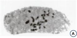
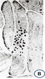
Figure 10.3: Cells from the root-tips of shoots of endosperm origin: A. showing diploid number of chromosomes (2n=2x=24), B. triploid number of chromosomes (2n=3x=36)