4. Histology
Histological studies of the proliferating endosperm of Jatropa, Putranjiva and Ricinus, revealed that the embryo also enlarged and proliferated along with the endosperm but soon showed the sign of degeneration. In such cases the endosperm calli were transferred to a fresh medium to avoid any contamination from degenerated embryonal cells. The 4-week-old callus derived from endosperm cultures, proliferated into parenchymatous cells and 6-week old callus showed tracheidal cells (Srivastava 1971a, b; Srivastava 1973; Johri and Srivastava 1973). In Santalum, endosperm proliferation started after the formation of several meristematic layers below the epidermal region (Rangaswamy and Rao 1963). By carefully applying plant growth regulators the nodular outgrowths can be induced on the surface of the cultured endosperm as in case of Osyris wightians (Johri and Bhojwani 1965) and Putranjiva roxburghii (Srivastava 1973).
The importance of tracheidal differentiation in the callus of endosperm cultures is that it facilitates organogenic differentiation. In the families like Euphorbiaceae, Loranthaceae and Santalaceae, the endosperm tissues readily form tracheidal elements in cultures (Johri and Srivastava 1973, Johri and Bhojwani 1971). In Emblica officinale, tracheidal cells and cambium like cells organized into vascular strands or nodules in the differentiation medium while in the callusing medium tracheidal cells remained scattered. The differentiation of vascular strand in the callus accompanied the shoot bud formation (Sehgal and Khurana 1985).
In Aleuritus fordii, callus proliferated from endosperm explant consisted of large, compact and vacuolated cells. Tiny group of cells became distinct from adjoining large and vacuolated cells and became meristematic. These cells remained thin walled with dense cytoplasm and a large clear nucleus. Later the meristemoids developed in to dome shaped shoot apex, which produces leaf primordia (Syed Abbas 1993). In Mallotus phillippensis only the compact green callus underwent differentiation. Such callus showed vasculature, developed protuberances and eventually gave rise to shoots buds. Small group of cells with deep-seated distinct meristematic loci were also observed in these calli, which later gave rise to dome-shaped shoot primordia, endogenously (Sehgal and Abbas 1996).
In mulberry (Morus alba), histological analysis revealed that the older region of the callus comprised of highly vacuolated cells. Shoot buds differentiated from peripheral nodular structures, which comprised of compactly arranged highly cytoplasmic cells. Often a few layers of degenerating vacuolated cells were seen outside the shoot primordia. It is possible that the shoots originated from inside the nodules and emerged after rupturing the surrounding tissue. The regenerating shoots showed vascular supply continuous with the vasculature of the callus (Thomas 2000).
Both exogenous and endogenous differentiation of shoots was observed in Azadirachta indica . The serial section of two-week-old regenerating callus showed that many meristematic pockets developed from inside the callus, which developed into shoot buds after 3 weeks (Figure 10.2A). Histological sections also revealed that the shoot buds emerged from the peripheral tissues of the callus as well (Chaturvedi et al. 2003) (Figure 10.2B). In Actinidia deliciosa histological analysis of the freshly isolated endosperm revealed small intercellular spaces and cells were filled with storage materials. However, the calli derived from the endosperm were larger, vacuolated and lacked storage materials. In older callus daughter nuclei attached to newly formed cell walls were often observed, suggesting disturbances of cell division. The cells differed in size and shape and contained nuclei with variable numbers of nucleoli (Goralski et al. 2005).
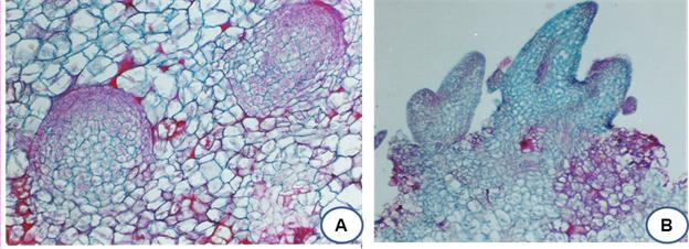
Figure 10.2: A. Section of a 2-week-old regenerating callus from endosperm of A. indica showing endogenous meristematic pockets. B. Section of a 4-week-old regenerating callus showing distinct shoot buds differentiated from peripheral vascularized nodule. One of the buds showing glands.