Fluorescence Microscope :
Some molecules absorb radiant energy, and become excited and later release much of their trapped energy as light, which has a longer wavelength and can be seen by the use of special light filters. The specimen to be viewed is stained with one of group of fluorescent dyes called fluorochromes. Then microorganism are examined under a fluorescent microscope with an UV or near UV light source, they appear as luminescent, bright objects against a dark background (Fig. 6). It has become essential tool in medical microbiology and microbial ecology. Fluorochromeauramine O glows yellow when exposed to UV light. This dye is strongly absorbed by the Mycobacterium tuberculosis and can be detected by the appearance of bright – yellow color. Bacillus anthracis , appears apple-green when stained with fluorescein isothiocyanate (FITC). Other fluorochromes are acridine orange, DAPI (diamidino-2-phenylindole, a DNA specific stain).
Principal use of FM is a diagnostic technique called the fluorescent-antibody technique or immunofluorescence. It is especially useful in diagnosing Syphilis and rabies. Antibodies – are natural defense molecules that are produced by humans and many animals in reaction to a foreign substance called antigen . Fluorescent antibodies for a particular antigen are obtained as follows:
• An animal is injected with a specific antigen, such as bacterium
• The animal then begins to produce specific antibodies
• After sufficient time, the antibiotics are removed from serum of the animal
• A fluorochrome is chemically combined with the antibodies
• These fluorescent antibodies are then added to a microscopic slide containing an unknown bacterium
• If this unknown is the same that was injected into the animal, the fluorescent antibodies bind to the antigens on the surface of the bacterium, causing it to fluoresce when observed under the fluorescence microscope
• This technique can detect bacteria or other disease producing microorganism even within cells, tissues or other clinical specimens
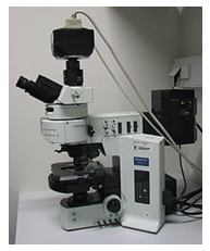
Fig. 6. Fluorescence microscope
Inverted microscopy:
The normal light microscope is functionally turned upside down. It is useful in tissue culture since it allows observation of cells through the bottom of a culture vessel, without opening the container, and without the air interface normally present between the objective and the surface of the culture (Fig. 7). By adding phase contrast optics to the inverted microscope, it is possible to monitor tissue cultures directly, without the aid of stains or other enhancements.
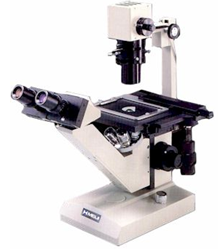
Fig. 7. Inverted microscope
REFERENCES:
Text Books:
1. Jeffery C. Pommerville. Alcamo's Fundamentals of Microbiology (Tenth Edition). Jones and Bartlett Student edition.
2. Gerard J. Tortora, Berdell R. Funke, Christine L. Case. Pearson - Microbiology: An Introduction. Benjamin Cummings.
Reference Books:
1. Lansing M. Prescott, John P. Harley and Donald A. Klein. Microbiology. Mc Graw Hill companies.