Bright field Microscope:
Bright field microscopy is the simplest of all the optical microscopy illumination techniques (Fig. 2). Sample illumination is transmitted (i.e., illuminated from below and observed from above) white light and contrast in the sample is caused by absorbance of some of the transmitted light in dense areas of the sample. The typical appearance of a bright field microscopy image is a dark sample on a bright background. Nonviable, stained preparations can be observed. Maximum resolution (ability of lens to separate or distinguish between small objects that are close together) of a light microscope is about 0.2 m m. The resolution can be improved with a sub stage condenser which focuses a cone of light on the specimen. Simplicity of setup with only basic equipment required is the main advantage. The main disadvantages are:- very low contrast of most biological samples, low apparent optical resolution due to the blur of out of focus material, the sample often has to be stained before viewing. Therefore, live cells cannot usually be viewed.
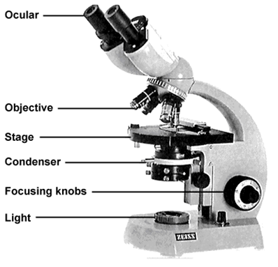
Fig. 2. Bright field microscope
Dark field microscope:
Living, unstained cells and organisms can be observed by changing the way in which they are illuminated. A dark field condenser that contains an opaque disc is used. The disc blocks light that would enter the objective directly. Only light that is diffracted by the specimen enters the objective lens (Fig. 3). The field surrounding a specimen appears black, while the object itself is brightly illuminated. It is based on light scattered at boundaries between regions having different refractive indexes. It is also used to identify bacteria like the thin and distinctively shaped Treponemapallidum, the causative agent of syphilis.
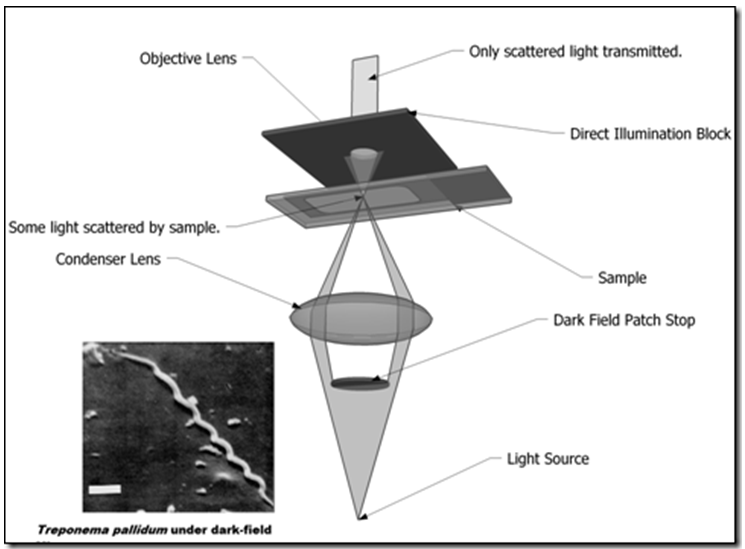
Fig. 3. Dark filed microscopy
Phase contrast microscope :
Especially useful for studying internal structures in living microorganisms and also studying microbial motility, determining the shape of living cells and detecting bacterial components such as endospores and inclusion bodies. Phase-contrast microscopy uses diffraction plate to diffract light rays so that they are out phase with one another; the specimen appears different degree of brightness and contrast (Fig. 4). It is based on the principle that when a light passing through an object undergo a phase change. Converts variations in the refractive index and density of cells into changes in light intensity and thus makes colorless, unstained cells visible.
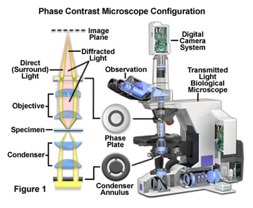
Fig. 4. Phase contrast microscope
Differential interference contrast microscope:
Similar to phase contrast and uses two beams of light to create high-contrast, three-dimensional images of live specimens. Structures such as cell walls, endospores, granules, vacuoles and eukaryotic nuclei are clearly visible. Differential interference contrast microscope uses difference in refractive indexes to produce image and are superb for both observation and measuring thickness of embryos within specimens with little or no contrast (Fig. 5).
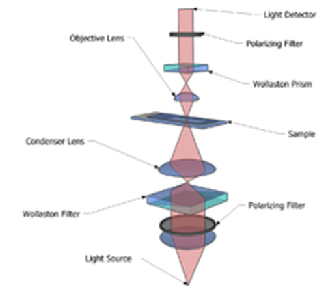
Fig. 5. The components of the basic differential interference contrast microscope setup.